Copyright
©The Author(s) 2016.
World J Stem Cells. Dec 26, 2016; 8(12): 399-427
Published online Dec 26, 2016. doi: 10.4252/wjsc.v8.i12.399
Published online Dec 26, 2016. doi: 10.4252/wjsc.v8.i12.399
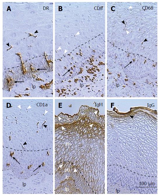
Figure 2 Distribution of monocyte derived cells, T cells, and immunoglobulins in the squamous epithelium of uterine ectocervix.
A: Dendritic cell (DC) precursors accompany vessels in the lamina propria (lp), release HLA-DR in the parabasal layer (arrows), and form DC (arrowheads) in the intermediate layer; B: T cells associate with basal epithelial cells, reach parabasal(arrow)/intermediate interface, and degenerate thereafter (arrowheads); C: The CD68 expression (arrow) accompanies mature DC (black arrowheads), which secrete CD68 (white arrowheads); D: CD1a+ DC precursors (arrows) and mature DC (black arrowheads) undergoing fragmentation (white arrowheads); E: The IgM binds to upper parabasal, upper intermediate, and upper superficial layers (arrowheads); F: IgG binds to the entire superficial layer only. The lp indicates epithelium lp, doted line is basement membrane, and dashed line is parabasal/intermediate interface[14].
- Citation: Bukovsky A. Involvement of blood mononuclear cells in the infertility, age-associated diseases and cancer treatment. World J Stem Cells 2016; 8(12): 399-427
- URL: https://www.wjgnet.com/1948-0210/full/v8/i12/399.htm
- DOI: https://dx.doi.org/10.4252/wjsc.v8.i12.399


