Copyright
©The Author(s) 2015.
World J Stem Cells. Aug 26, 2015; 7(7): 1047-1053
Published online Aug 26, 2015. doi: 10.4252/wjsc.v7.i7.1047
Published online Aug 26, 2015. doi: 10.4252/wjsc.v7.i7.1047
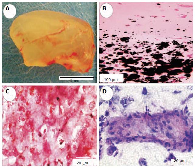
Figure 2 Engineered neogenesis of human-shaped mandibular condyle from mesenchymal stem cells.
A: Harvested osteochondral construct retained the shape and dimension of the cadaver human mandibular condyle after in vivo implantation; B: Von Kossa-stained section showing the interface between stratified chondral and osseous layers. Multiple mineralization nodules are present in the osseous layer (lower half of the photomicrograph), but absent in the chondral layer; C: Positive safranin O staining of the chondrogenic layer indicates the synthesis of abundant glycosaminoglycans; D: H and E-stained section of the osteogenic layer showing a representative osseous island-like structure consisting of MSC-differentiated osteoblast-like cells on the surface and in the center. Reproduced with permission from Biomedical Engineering Society[36].
- Citation: Aly LAA. Stem cells: Sources, and regenerative therapies in dental research and practice. World J Stem Cells 2015; 7(7): 1047-1053
- URL: https://www.wjgnet.com/1948-0210/full/v7/i7/1047.htm
- DOI: https://dx.doi.org/10.4252/wjsc.v7.i7.1047


