Copyright
©The Author(s) 2015.
World J Stem Cells. Jul 26, 2015; 7(6): 922-944
Published online Jul 26, 2015. doi: 10.4252/wjsc.v7.i6.922
Published online Jul 26, 2015. doi: 10.4252/wjsc.v7.i6.922
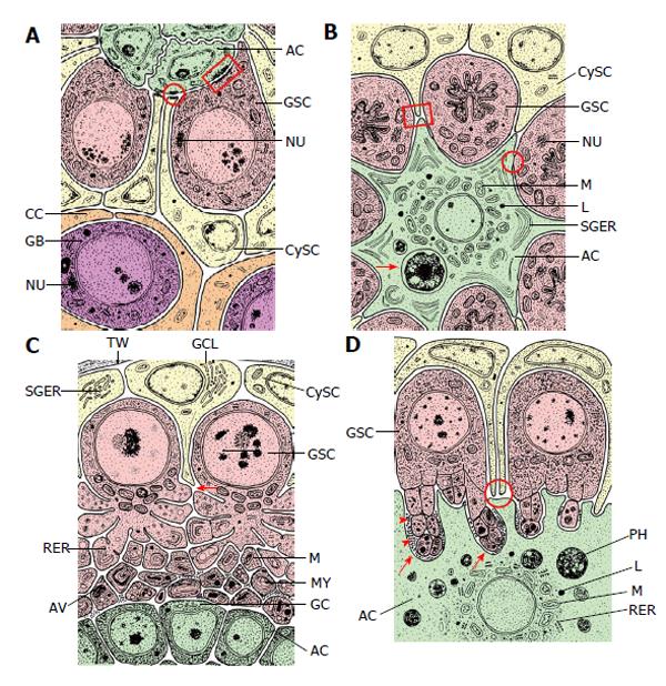
Figure 5 Fine structural organization (schematized) of the apical complexes (longitudinal sections) of four insect species.
The order (A) to (D) is arranged according to the order in Figure 3. A: Drosophila melanogaster. ACs are “light” and interdigitate richly between themselves. Their organelle equipment is scarce. GSCs are “dark” due to many free ribosomes and characterized by “spongy bodies” which may represent nuage. Adherens junctions are expressed between ACs and GSCs (marked by a rectangle) and between ACs and CySCs (encircled). CySCs are “light” and include few cell organelles (Adapted from Hardy et al[61]); B: Locusta migratoria. The single apical cell of an apical complex is “light” and harbors a complex organelle equipment: in particular a ring of mitochondria (M) and lysosomes (L) around the nucleus, and stacks of sparsely granulated endoplasmic reticulum (SGER) at the cell periphery. Arrow marks a phagocytized and partly lysed GSC. ACs and GSCs are connected by gap-like junctions (encircled). The “dark” GSCs include extremely irregularly shaped nuclei and nuage-like material (NU). Extensions of the “light” CySCs reach to extensions of the star-shaped AC (marked by square) (Adapted from Dorn et al[64]); C: Oncopeltus fasciatus. The ACs are rather small and very “dark”. Cell organelles are inconspicuous besides Golgi complexes (GC) that face bordering GSC vesicles. The “dark” GSCs form cytoplasmic projections toward the ACs that undergo progressing autotomy. In the course of vesicle segregation rough endoplasmic reticulum (RER) and mitochondria (M) increase. Later autophagic vacuoles (AV) and myelin-like bodies (MY) are formed. Arrow points to a presumably newly sprouting cell projection. The “light” CySCs are characterized by extensive Golgi complex-like structures (GCL) that bare often associated with sparsely granulated endoplasmic reticulum (SGER) (Adapted from Dorn et al[60]); D: Lymantria dispar. The single AC of an apical complex is “light” and exhibits a spherical organization: mitochondria (M), lysosomes (L) and rough endoplasmic reticulum (RER) surround the nucleus: The cell periphery shows deep indentations caused by invading, autotomizing GSC projections (arrows). Segregated GSC vesicles are taken up by the AC, and phagosomes (PH) accumulate in the cytoplasm. The “dense” GSCs exhibit projection formation and projection autotomy that closely resembles that of Oncopeltus (see Figure 4C). GSC projections that indent the AC are often surrounded by extracellular granules (arrow heads). CySCs are “light”, and thin extensions reach the surface of the GSCs (encircled) (Adapted from Klein[66]). AC: Apical cell (green); CC: Cyst cell (orange); CySC: Cyst stem cell (yellow); GB: Gonialblast (purple); GSC: Germline stem cell (red); TW: Testicular wall (grey); AV: Autophagic vacuole; GC: Golgi complex; GCL: Golgi complex-like stricture; L: Lysosomes; M: Mitochondrion; MY: Myelin-like body; NU: Nuage; PH: Phagosomes; RER: Rough endoplasmic reticulum; SGER: Sparsely granulated endoplasmic reticulum.
- Citation: Dorn DC, Dorn A. Stem cell autotomy and niche interaction in different systems. World J Stem Cells 2015; 7(6): 922-944
- URL: https://www.wjgnet.com/1948-0210/full/v7/i6/922.htm
- DOI: https://dx.doi.org/10.4252/wjsc.v7.i6.922


