Copyright
©The Author(s) 2015.
World J Stem Cells. Mar 26, 2015; 7(2): 281-299
Published online Mar 26, 2015. doi: 10.4252/wjsc.v7.i2.281
Published online Mar 26, 2015. doi: 10.4252/wjsc.v7.i2.281
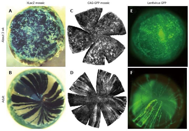
Figure 4 Transition from randomly orientated patches to radial stripes in corneal epithelia of different types of mosaic mice between 3 wk and adulthood.
A and B: β-gal staining in XLacZ X-inactivation mosaics[27]; C and D: Green fluorescent protein (GFP) fluorescence in CAG-GFP transgenic mosaics[77]; E and F: GFP fluorescence in corneal epithelium after transfecting conceptuses with lentiviral vectors encoding GFP at embryonic day 9 or 10[79]. Photographs (A and D) are reproduced from Developmental Dynamics[27] with kind permission of John Wiley and Sons, (C and D) are reproduced from Molecular Vision[77] with kind permission of the authors and editors, and photographs (E and F) are reproduced from Molecular Therapy[79] with kind permission of the authors and the Nature Publishing Group. This combination of photographs was first published by Mort et al[18].
- Citation: West JD, Dorà NJ, Collinson JM. Evaluating alternative stem cell hypotheses for adult corneal epithelial maintenance. World J Stem Cells 2015; 7(2): 281-299
- URL: https://www.wjgnet.com/1948-0210/full/v7/i2/281.htm
- DOI: https://dx.doi.org/10.4252/wjsc.v7.i2.281


