Copyright
©The Author(s) 2015.
World J Stem Cells. Mar 26, 2015; 7(2): 266-280
Published online Mar 26, 2015. doi: 10.4252/wjsc.v7.i2.266
Published online Mar 26, 2015. doi: 10.4252/wjsc.v7.i2.266
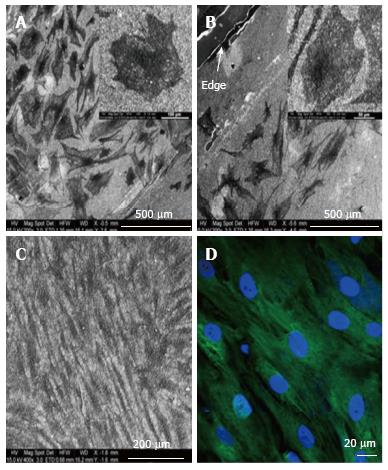
Figure 8 Electron microscopy images of differentiated and undifferentiated mesenchymal stem cells on Coll/PLLCL nanostructured nanofibrers.
A: Mesenchymal stem cell (MSC) directed to differentiate along the epidermal lineage when cultured in epidermal induction medium; B: Epidermally differentiated MSC on the edge of a Coll/PLLCL scaffold (as shown by arrow); C: Electrospun Coll/PLLCL nanofibers seeded with undifferentiated MSC cultured in normal growth medium; D: MSC grown in normal growth medium on Coll/PLLCL nanofibers stained with Ker 10, after 15 d cell culture, imaged using laser scanning confocal microscope[85].
- Citation: Salmasi S, Kalaskar DM, Yoon WW, Blunn GW, Seifalian AM. Role of nanotopography in the development of tissue engineered 3D organs and tissues using mesenchymal stem cells. World J Stem Cells 2015; 7(2): 266-280
- URL: https://www.wjgnet.com/1948-0210/full/v7/i2/266.htm
- DOI: https://dx.doi.org/10.4252/wjsc.v7.i2.266


