Copyright
©The Author(s) 2015.
World J Stem Cells. Mar 26, 2015; 7(2): 266-280
Published online Mar 26, 2015. doi: 10.4252/wjsc.v7.i2.266
Published online Mar 26, 2015. doi: 10.4252/wjsc.v7.i2.266
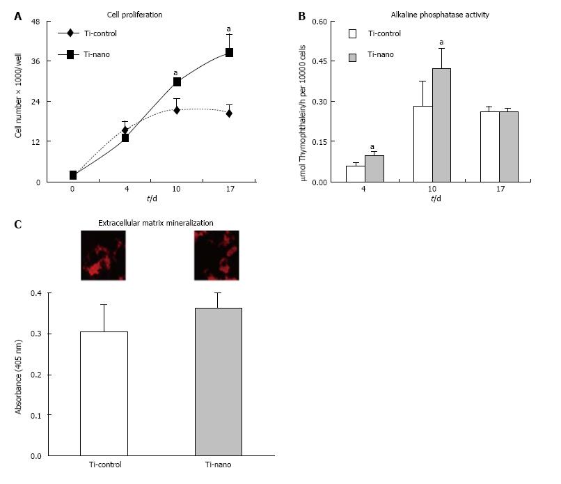
Figure 6 Investigation of the effects of nanotopography on proliferation.
(A), alkaline phosphatase (Alp) activity (B), and extracellular matrix mineralization (C) of mesenchymal stem cells differentiated into osteoblasts and cultured on nanotopography in an osteogenic medium compared to control Ti surfaces. A: The number of cells was significantly increased on Ti with nanotopography on days 10 (P = 0.07) and 17 (P = 0.03); B: Higher Alp activity was supported by Ti surface with nanotopography supported on days 4 (P = 0.01) and 10 (P = 0.04); C: The difference in the level of calcium mineralisation in the matrix (insets) was not statistically significant (P = 0.13) by comparing both surfaces. The data are presented as mean ± standard deviation (n = 4). aIndicates statistically significant difference[66].
- Citation: Salmasi S, Kalaskar DM, Yoon WW, Blunn GW, Seifalian AM. Role of nanotopography in the development of tissue engineered 3D organs and tissues using mesenchymal stem cells. World J Stem Cells 2015; 7(2): 266-280
- URL: https://www.wjgnet.com/1948-0210/full/v7/i2/266.htm
- DOI: https://dx.doi.org/10.4252/wjsc.v7.i2.266


