Copyright
©The Author(s) 2015.
World J Stem Cells. Mar 26, 2015; 7(2): 266-280
Published online Mar 26, 2015. doi: 10.4252/wjsc.v7.i2.266
Published online Mar 26, 2015. doi: 10.4252/wjsc.v7.i2.266
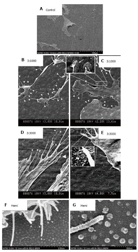
Figure 5 Scanning electron micrographs of human mesenchymal stem cells cultured on control and test materials.
A: On planar control materials cells showed normal morphologies; B, C: Filopodial of the human mesenchymal stem cells (hMSCs) interacts with the 3:1000 substrates (arrowheads); C: hMSC filopodia are curving around an island; D, E: Filopodial interactions with the 3:3000 substrates (arrowheads); E: A filopodia curving around an island is clearly observed; F: filopodial interactions with the hemi substrates (arrowheads); G: Filopodia curving around a hemisphere (arrowhead)[65].
- Citation: Salmasi S, Kalaskar DM, Yoon WW, Blunn GW, Seifalian AM. Role of nanotopography in the development of tissue engineered 3D organs and tissues using mesenchymal stem cells. World J Stem Cells 2015; 7(2): 266-280
- URL: https://www.wjgnet.com/1948-0210/full/v7/i2/266.htm
- DOI: https://dx.doi.org/10.4252/wjsc.v7.i2.266


