Copyright
©The Author(s) 2015.
World J Stem Cells. Mar 26, 2015; 7(2): 266-280
Published online Mar 26, 2015. doi: 10.4252/wjsc.v7.i2.266
Published online Mar 26, 2015. doi: 10.4252/wjsc.v7.i2.266
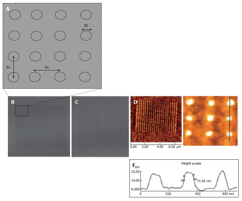
Figure 4 Nanopatterned gold surfaces examination for the effect of both the nanotopography and terminating chemical functionality.
A: Nanopatterned surfaces used for mesenchymal stem cell control and differentiation exhibiting dot to dot pitch (dα) and dot diameter (dβ); B: Lateral Force Microscopy (LFM) image of small area 280 nm pitch array; C: LFM image of 140 nm pitch array; D-F: Atomic force microscopy topographical image of an alkanethiol resist array fabricated on gold surface following chemical etching. An average diameter feature (dβ) of 70 nm was shown on the cursor profile[49].
- Citation: Salmasi S, Kalaskar DM, Yoon WW, Blunn GW, Seifalian AM. Role of nanotopography in the development of tissue engineered 3D organs and tissues using mesenchymal stem cells. World J Stem Cells 2015; 7(2): 266-280
- URL: https://www.wjgnet.com/1948-0210/full/v7/i2/266.htm
- DOI: https://dx.doi.org/10.4252/wjsc.v7.i2.266


