Copyright
©The Author(s) 2015.
World J Stem Cells. Dec 26, 2015; 7(11): 1251-1261
Published online Dec 26, 2015. doi: 10.4252/wjsc.v7.i11.1251
Published online Dec 26, 2015. doi: 10.4252/wjsc.v7.i11.1251
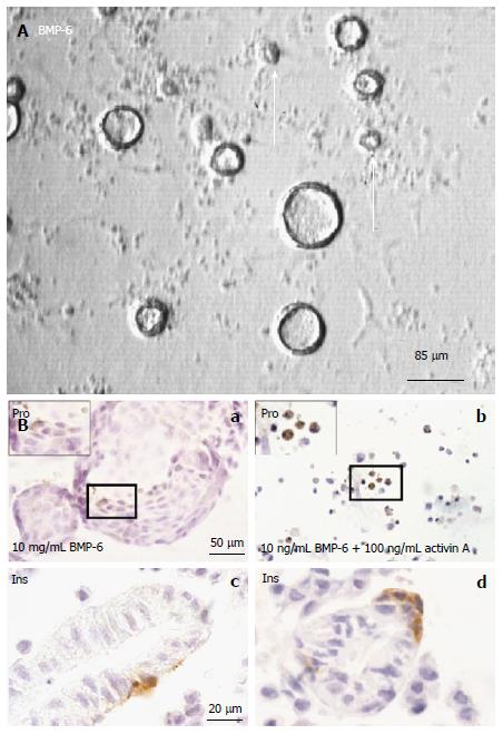
Figure 2 Cystic colony formation from dissociated fetal mouse pancreas cells.
A: Phase contrast image showing that BMP-6 promotes colony formation. Open arrows indicate colonies ≤ 30 μm; B: Immunocytochemical analyses: a: Proinsulin staining. Fixed colonies were stained with proinsulin antibody (brown); b: Activin A antagonizes colony formation; c, d: Insulin staining. Histological sections of harvested colonies were stained with anti-insulin antibody (brown). Adapted and modified from ref.[14].
- Citation: Jiang FX, Morahan G. Multipotent pancreas progenitors: Inconclusive but pivotal topic. World J Stem Cells 2015; 7(11): 1251-1261
- URL: https://www.wjgnet.com/1948-0210/full/v7/i11/1251.htm
- DOI: https://dx.doi.org/10.4252/wjsc.v7.i11.1251


