Copyright
©The Author(s) 2015.
World J Stem Cells. Jan 26, 2015; 7(1): 75-83
Published online Jan 26, 2015. doi: 10.4252/wjsc.v7.i1.75
Published online Jan 26, 2015. doi: 10.4252/wjsc.v7.i1.75
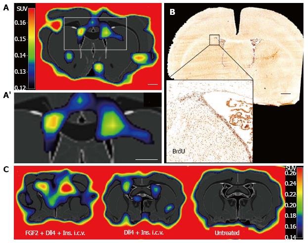
Figure 2 [18F]FLT labels proliferating endogenous neural stem cell in the neurogenic niches of the healthy rodent brain.
A, A’: eNSC proliferation in the subventricular zone of adult rats as assessed by [18F]FLT-PET; B: The signal corresponds to BrdU-positive cells in the region; C: Activation of eNSC by pharmacological stimulation with fibroblast growth factor 2, delta-like 4, and insulin is visualized with [18F]FLT-PET. eNSC: Endogenous neural stem cell; [18F]FLT-PET: 3’-deoxy-3’-[18F]fluoro-L-thymidine-positron emission tomography; i.c.v.: Intracerebroventricular application. Adapted from Rueger et al[75], with permission.
-
Citation: Rueger MA, Schroeter M.
In vivo imaging of endogenous neural stem cells in the adult brain. World J Stem Cells 2015; 7(1): 75-83 - URL: https://www.wjgnet.com/1948-0210/full/v7/i1/75.htm
- DOI: https://dx.doi.org/10.4252/wjsc.v7.i1.75


