Copyright
©The Author(s) 2018.
World J Stem Cells. Jan 26, 2018; 10(1): 1-14
Published online Jan 26, 2018. doi: 10.4252/wjsc.v10.i1.1
Published online Jan 26, 2018. doi: 10.4252/wjsc.v10.i1.1
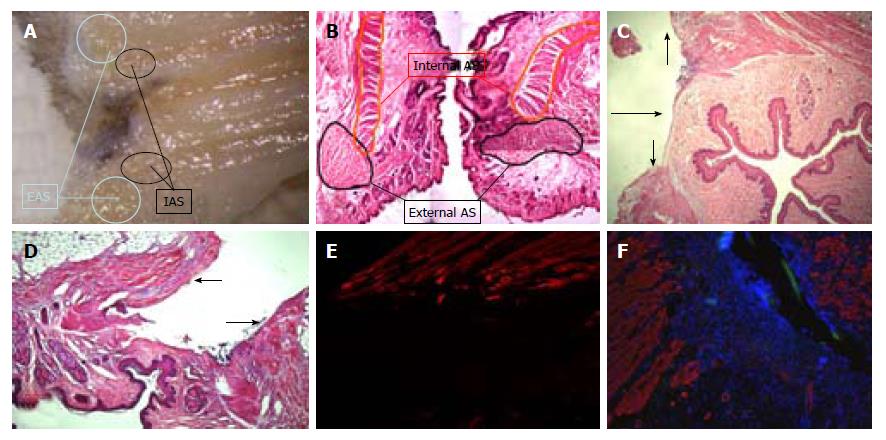
Figure 5 Anatomy and injury model study.
Sometimes it is difficult to identify the external anal sphincter (see Figure 2), but it is present and included in the section as can be seen in A: A sagittal section; or in B: Coronal hematoxylin and eosin reconstruction (10 ×). The described model implies section of internal anal sphincter (C: Hematoxylin and eosin 10 ×) and external anal sphincter (D: Hematoxylin and eosin 10 ×). This was also evidenced on immunofluorescence staining at 10 × (E belongs to a non-repaired rat and F to a biosuture group rat; suture strains can be seen with green autofluorescence). EAS: External anal sphincter; IAS: Internal anal sphincter; AS: Anal sphincter.
- Citation: Trébol J, Georgiev-Hristov T, Vega-Clemente L, García-Gómez I, Carabias-Orgaz A, García-Arranz M, García-Olmo D. Rat model of anal sphincter injury and two approaches for stem cell administration. World J Stem Cells 2018; 10(1): 1-14
- URL: https://www.wjgnet.com/1948-0210/full/v10/i1/1.htm
- DOI: https://dx.doi.org/10.4252/wjsc.v10.i1.1


