Copyright
©The Author(s) 2003.
World J Gastroenterol. Apr 15, 2003; 9(4): 645-649
Published online Apr 15, 2003. doi: 10.3748/wjg.v9.i4.645
Published online Apr 15, 2003. doi: 10.3748/wjg.v9.i4.645
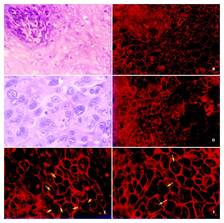
Figure 2 The translocation of annexin I protein in ESCC.
Paraffin-embedded tissue sections of ESCC were detected with anti-annexin I antibody and annexin I protein was labeled by TRITC-conjugated secondary antibody. The fluorescence image was visualized under a fluorescence microscope (Olympus BX51, Japan). An H & E staining was performed (a 200 ×; c 400 ×). A hyperplasia of esophageal epithelia were shown in Figure 2 (a and b, b 200 ×) and there were basal papillae displaying bound-ary area of the epithelium, which consecutively expressed annexin I formed the typical trammel net on the cellular membrane. Annexin I protein was decreased sharply and translocated from cellular membrane to the nuclear membrane (d 200 ×; e 400 ×; f 400 ×). The yellow arrows showed the nuclear membrane localization of annexin I in ESCC and all of the materials were from a same ESCC case.
- Citation: Liu Y, Wang HX, Lu N, Mao YS, Liu F, Wang Y, Zhang HR, Wang K, Wu M, Zhao XH. Translocation of annexin I from cellular membrane to the nuclear membrane in human esophageal squamous cell carcinoma. World J Gastroenterol 2003; 9(4): 645-649
- URL: https://www.wjgnet.com/1007-9327/full/v9/i4/645.htm
- DOI: https://dx.doi.org/10.3748/wjg.v9.i4.645


