Copyright
©The Author(s) 2023.
World J Gastroenterol. Mar 7, 2023; 29(9): 1427-1445
Published online Mar 7, 2023. doi: 10.3748/wjg.v29.i9.1427
Published online Mar 7, 2023. doi: 10.3748/wjg.v29.i9.1427
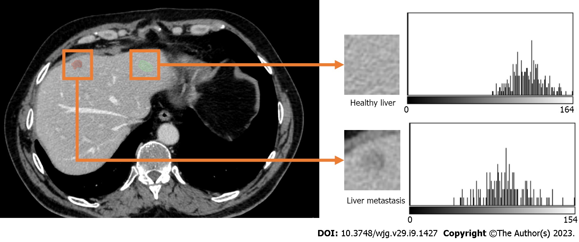
Figure 5 Computerized tomography scan of a 61-year-old male patient with colon carcinoma and liver metastases.
The intensity histograms of regions with and without metastases are different; hence, the first order radiomics features[109], which are based on the intensity histogram will potentially be different.
- Citation: Berbís MA, Paulano Godino F, Royuela del Val J, Alcalá Mata L, Luna A. Clinical impact of artificial intelligence-based solutions on imaging of the pancreas and liver. World J Gastroenterol 2023; 29(9): 1427-1445
- URL: https://www.wjgnet.com/1007-9327/full/v29/i9/1427.htm
- DOI: https://dx.doi.org/10.3748/wjg.v29.i9.1427


