Copyright
©The Author(s) 2023.
World J Gastroenterol. Sep 14, 2023; 29(34): 5038-5053
Published online Sep 14, 2023. doi: 10.3748/wjg.v29.i34.5038
Published online Sep 14, 2023. doi: 10.3748/wjg.v29.i34.5038
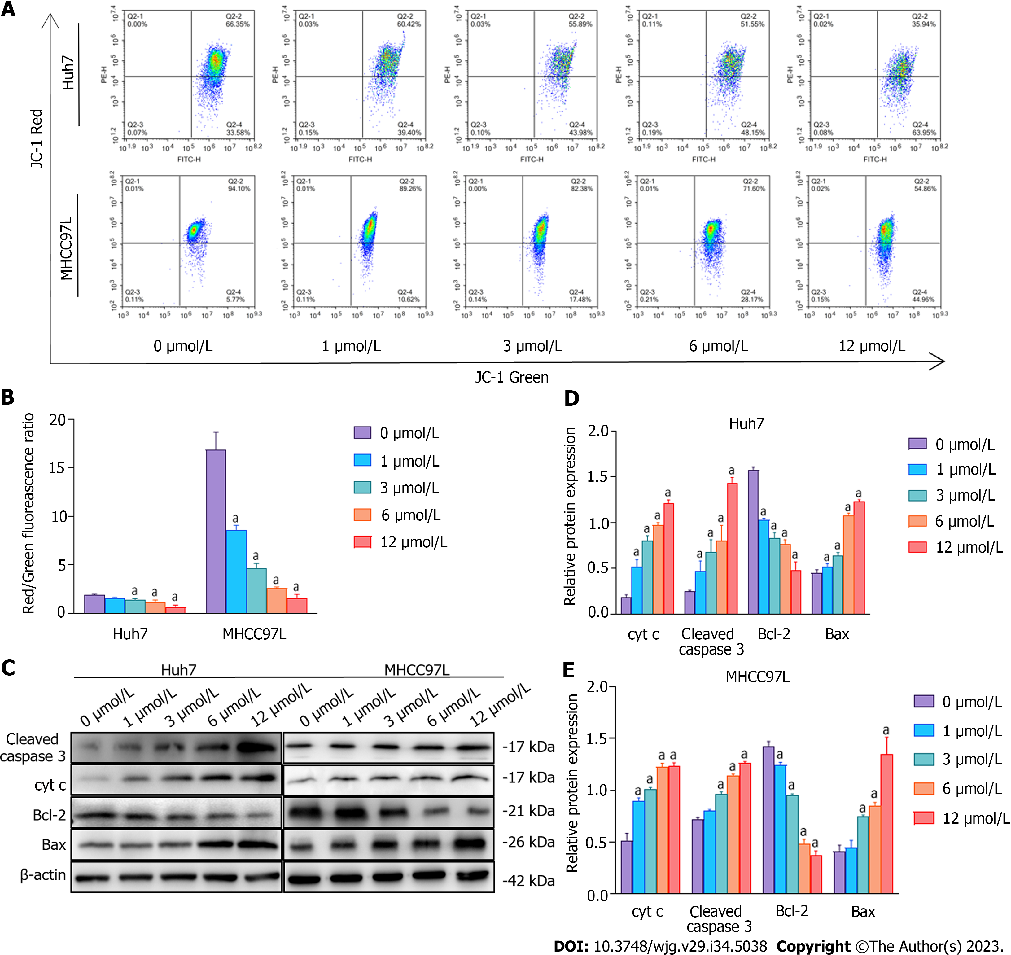
Figure 6 The impact of suberoylanilide hydroxamic acid treatment on mitochondrial membrane potential and the expression of proteins related to the mitochondrial apoptotic pathway in Huh7 and MHCC97L cells.
A and B: After being treated with 0, 1, 3, 6, and 12 μmol/L suberoylanilide hydroxamic acid for 48 h, the mitochondrial membrane potential in Huh7 and MHCC97L cells was analyzed using flow cytometry with bivariable JC-1 dye (mitochondrial membrane potential probe). Representative pictures from three independent replicate assays are exhibited; C-E: Representative western blot images showed the related protein expression in Huh7 and MHCC97L cells treated with 0, 1, 3, 6, and 12 μmol/L suberoylanilide hydroxamic acid for 48 h. Relative protein expression levels were normalized to β-actin. Data are exhibited as mean ± standard deviation. aP < 0.05 vs 0 μmol/L group (n = 3).
- Citation: Li JY, Tian T, Han B, Yang T, Guo YX, Wu JY, Chen YS, Yang Q, Xie RJ. Suberoylanilide hydroxamic acid upregulates reticulophagy receptor expression and promotes cell death in hepatocellular carcinoma cells. World J Gastroenterol 2023; 29(34): 5038-5053
- URL: https://www.wjgnet.com/1007-9327/full/v29/i34/5038.htm
- DOI: https://dx.doi.org/10.3748/wjg.v29.i34.5038


