Copyright
©The Author(s) 2023.
World J Gastroenterol. May 7, 2023; 29(17): 2534-2550
Published online May 7, 2023. doi: 10.3748/wjg.v29.i17.2534
Published online May 7, 2023. doi: 10.3748/wjg.v29.i17.2534
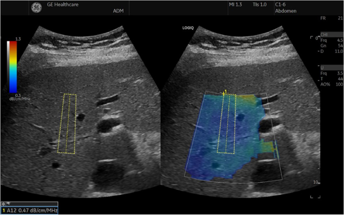
Figure 4 The ultrasound-guided attenuation parameter method implemented in the LOGIQ E9 XDclear 2.
0 US scanner[4]. The attenuation coefficient is 0.47 dB/cm/MHz, indicating less than 5% steatosis. Citation: Ferraioli G, Berzigotti A, Barr RG, Choi BI, Cui XW, Dong Y, Gilja OH, Lee JY, Lee DH, Moriyasu F, Piscaglia F, Sugimoto K, Wong GL, Wong VW, Dietrich CF. Quantification of Liver Fat Content with Ultrasound: A WFUMB Position Paper. Ultrasound Med Biol 2021; 47: 2803-2820. Copyright© The Author(s) 2021. Published by Elsevier Ltd. The authors have obtained the permission for figure using (Supplementary material).
- Citation: Zeng KY, Bao WYG, Wang YH, Liao M, Yang J, Huang JY, Lu Q. Non-invasive evaluation of liver steatosis with imaging modalities: New techniques and applications. World J Gastroenterol 2023; 29(17): 2534-2550
- URL: https://www.wjgnet.com/1007-9327/full/v29/i17/2534.htm
- DOI: https://dx.doi.org/10.3748/wjg.v29.i17.2534


