Copyright
©The Author(s) 2021.
World J Gastroenterol. Dec 14, 2021; 27(46): 7894-7908
Published online Dec 14, 2021. doi: 10.3748/wjg.v27.i46.7894
Published online Dec 14, 2021. doi: 10.3748/wjg.v27.i46.7894
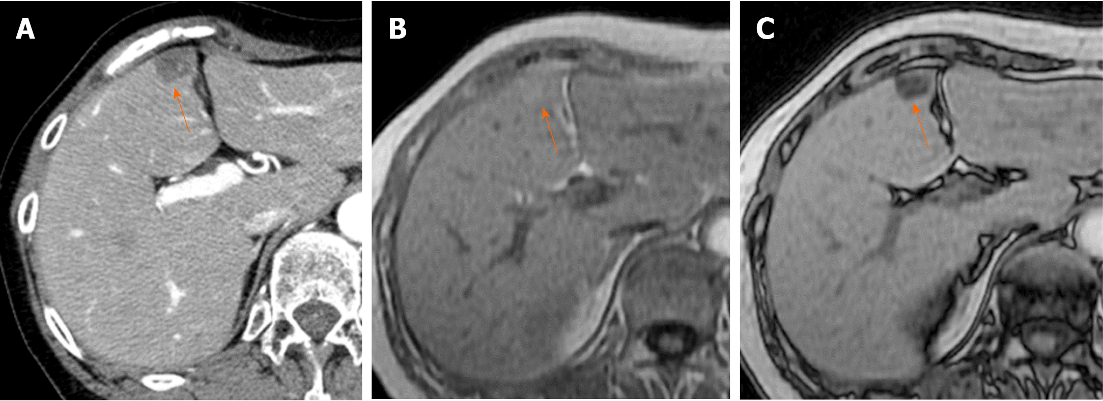
Figure 14 Focal fat deposition in the drainage area of the vein of Sappey (60th female).
A: On arterial phase contrast enhanced computed tomography image, focal hypoattenuation area is observed in anterior portion of segment IV of the liver adjacent to the falciform ligament (arrow). B and C: On T1 weighted in-phase and opposed-phase image of the liver, the lesion shows hyperintense on in-phase (B, arrow) and shows hypointense on opposed-phase (C, arrow), which represent focal fat deposition of the liver at the drainage area of inferior vein of Sappey.
- Citation: Kobayashi S. Hepatic pseudolesions caused by alterations in intrahepatic hemodynamics. World J Gastroenterol 2021; 27(46): 7894-7908
- URL: https://www.wjgnet.com/1007-9327/full/v27/i46/7894.htm
- DOI: https://dx.doi.org/10.3748/wjg.v27.i46.7894


