Copyright
©The Author(s) 2021.
World J Gastroenterol. Dec 14, 2021; 27(46): 7894-7908
Published online Dec 14, 2021. doi: 10.3748/wjg.v27.i46.7894
Published online Dec 14, 2021. doi: 10.3748/wjg.v27.i46.7894
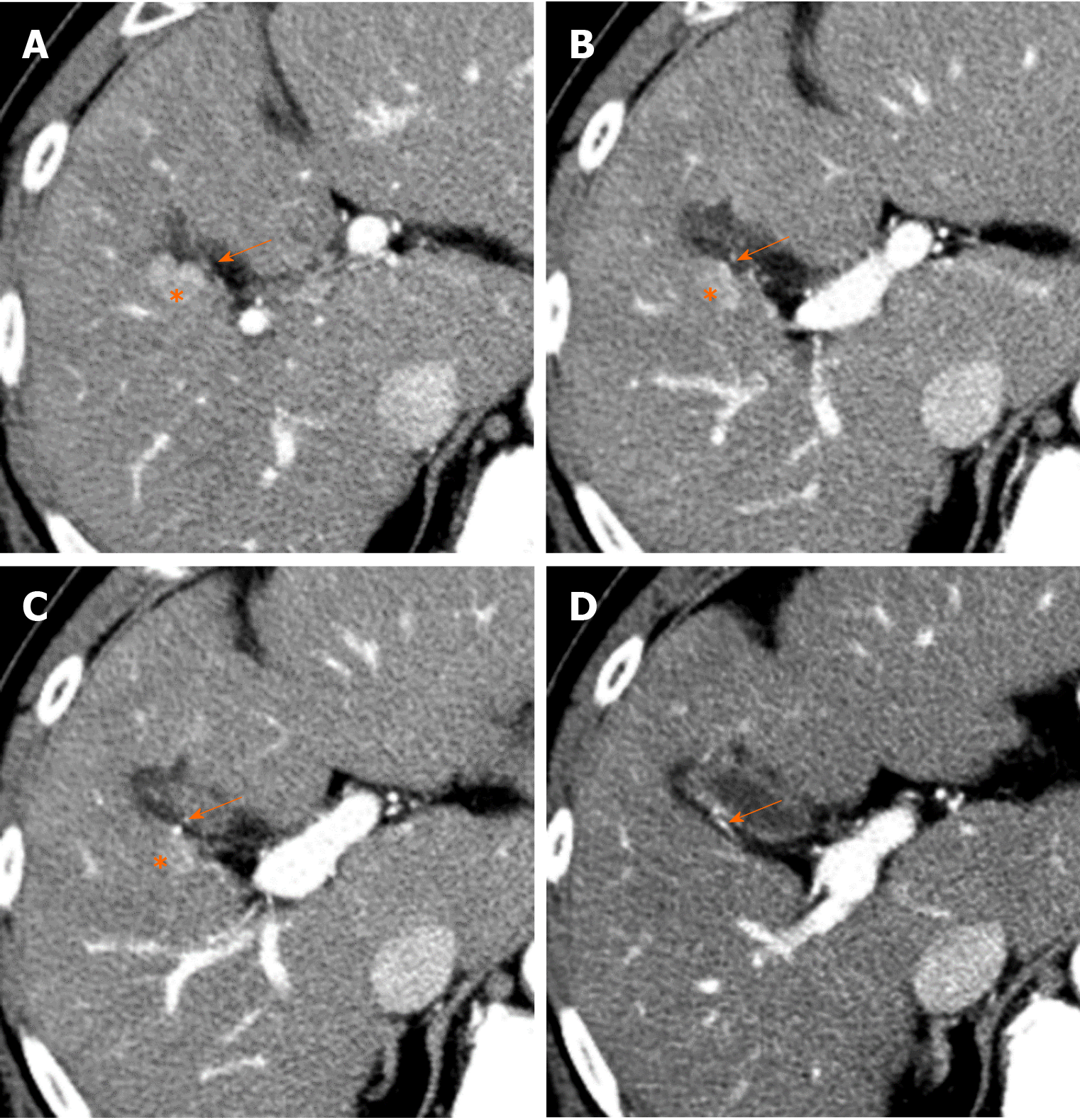
Figure 10 Hypervascular pseudolesion in cholecystic venous drainage area (50th male).
A-D: On sequential images of arterial phase contrast enhanced computed tomography, round hyper-attenuation area is observed in segment V of the liver adjacent to the gallbladder (*). Tiny enhanced vessel is directly entered to the enhanced liver area from the gallbladder wall (arrows), which is cholecystic venous drainage to the liver and hypervascular pseudolesion in cholecystic venous drainage area.
- Citation: Kobayashi S. Hepatic pseudolesions caused by alterations in intrahepatic hemodynamics. World J Gastroenterol 2021; 27(46): 7894-7908
- URL: https://www.wjgnet.com/1007-9327/full/v27/i46/7894.htm
- DOI: https://dx.doi.org/10.3748/wjg.v27.i46.7894


