Copyright
©The Author(s) 2020.
World J Gastroenterol. Sep 28, 2020; 26(36): 5450-5462
Published online Sep 28, 2020. doi: 10.3748/wjg.v26.i36.5450
Published online Sep 28, 2020. doi: 10.3748/wjg.v26.i36.5450
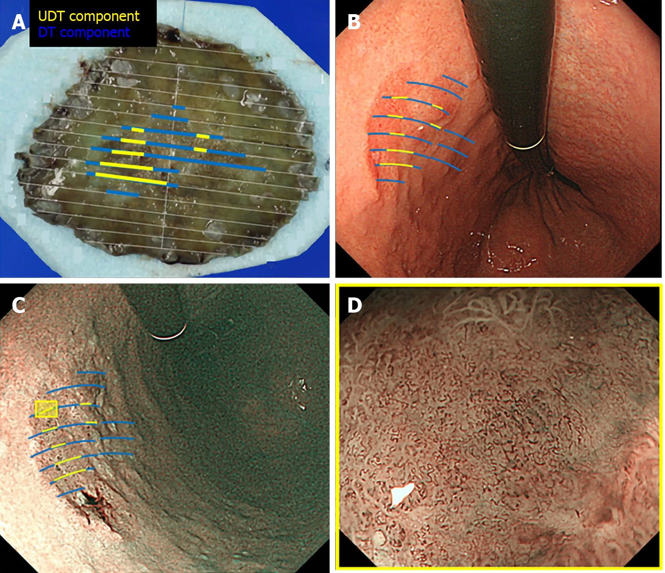
Figure 3 Correlation between pathology and endoscopic findings.
Preoperative endoscopic images were reviewed to determine whether the endoscopic finding of undifferentiated-type (UDT) component was recognizable within the area where UDT component was present in the histopathological examination. A: Endoscopically resected specimen cut into 2-mm-thick slices with mapping according to the histological types. Blue lines indicate differentiated-type component, while yellow lines indicate UDT component; B: Endoscopic images upon white light imaging onto which the histological type is mapped; C: Endoscopic images upon narrow-band imaging onto which the histological type is mapped; D: Magnifying endoscopy with narrow-band imaging finding of the designated UDT area with the yellow square in Figure 3C. ME-NBI: Magnifying endoscopy with narrow-band imaging; DT: Differentiated-type; UDT: Undifferentiated-type.
- Citation: Ozeki Y, Hirasawa K, Kobayashi R, Sato C, Tateishi Y, Sawada A, Ikeda R, Nishio M, Fukuchi T, Makazu M, Taguri M, Maeda S. Histopathological validation of magnifying endoscopy for diagnosis of mixed-histological-type early gastric cancer. World J Gastroenterol 2020; 26(36): 5450-5462
- URL: https://www.wjgnet.com/1007-9327/full/v26/i36/5450.htm
- DOI: https://dx.doi.org/10.3748/wjg.v26.i36.5450


