Copyright
©The Author(s) 2019.
World J Gastroenterol. Mar 28, 2019; 25(12): 1457-1464
Published online Mar 28, 2019. doi: 10.3748/wjg.v25.i12.1457
Published online Mar 28, 2019. doi: 10.3748/wjg.v25.i12.1457
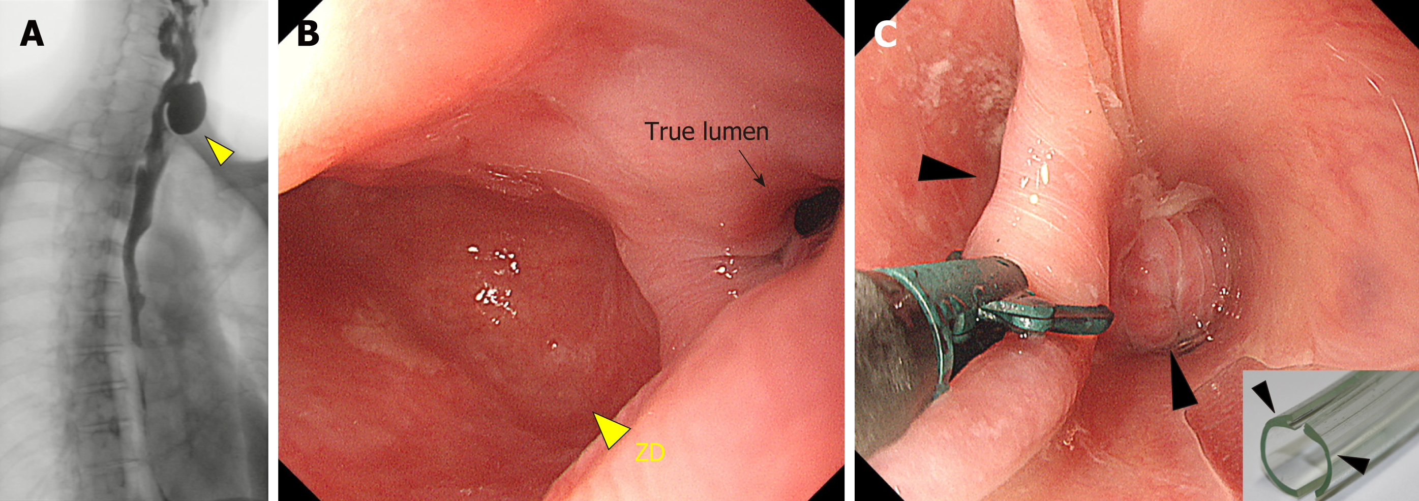
Figure 3 Zenker’s diverticulum treated using endoscopic diverticulectomy.
A: Zenker’s diverticulum (ZD, yellow triangle) visible on a barium swallow; B: On endoscopy, the ZD (yellow triangle) is easily visible and is bigger than the true esophageal lumen (black arrow); C: Endoscopic diverticulectomy is performed with a diverticuloscope (insert) straddled across the septum, with one flap inserted into the bottom of the ZD and the other in the esophageal lumen (black arrow) for a clear visualization of the septum and safe diverticulectomy[34]. ZD: Zenker’s diverticulum.
- Citation: Sato H, Takeuchi M, Hashimoto S, Mizuno KI, Furukawa K, Sato A, Yokoyama J, Terai S. Esophageal diverticulum: New perspectives in the era of minimally invasive endoscopic treatment. World J Gastroenterol 2019; 25(12): 1457-1464
- URL: https://www.wjgnet.com/1007-9327/full/v25/i12/1457.htm
- DOI: https://dx.doi.org/10.3748/wjg.v25.i12.1457


