Copyright
©The Author(s) 2018.
World J Gastroenterol. Jul 14, 2018; 24(26): 2806-2817
Published online Jul 14, 2018. doi: 10.3748/wjg.v24.i26.2806
Published online Jul 14, 2018. doi: 10.3748/wjg.v24.i26.2806
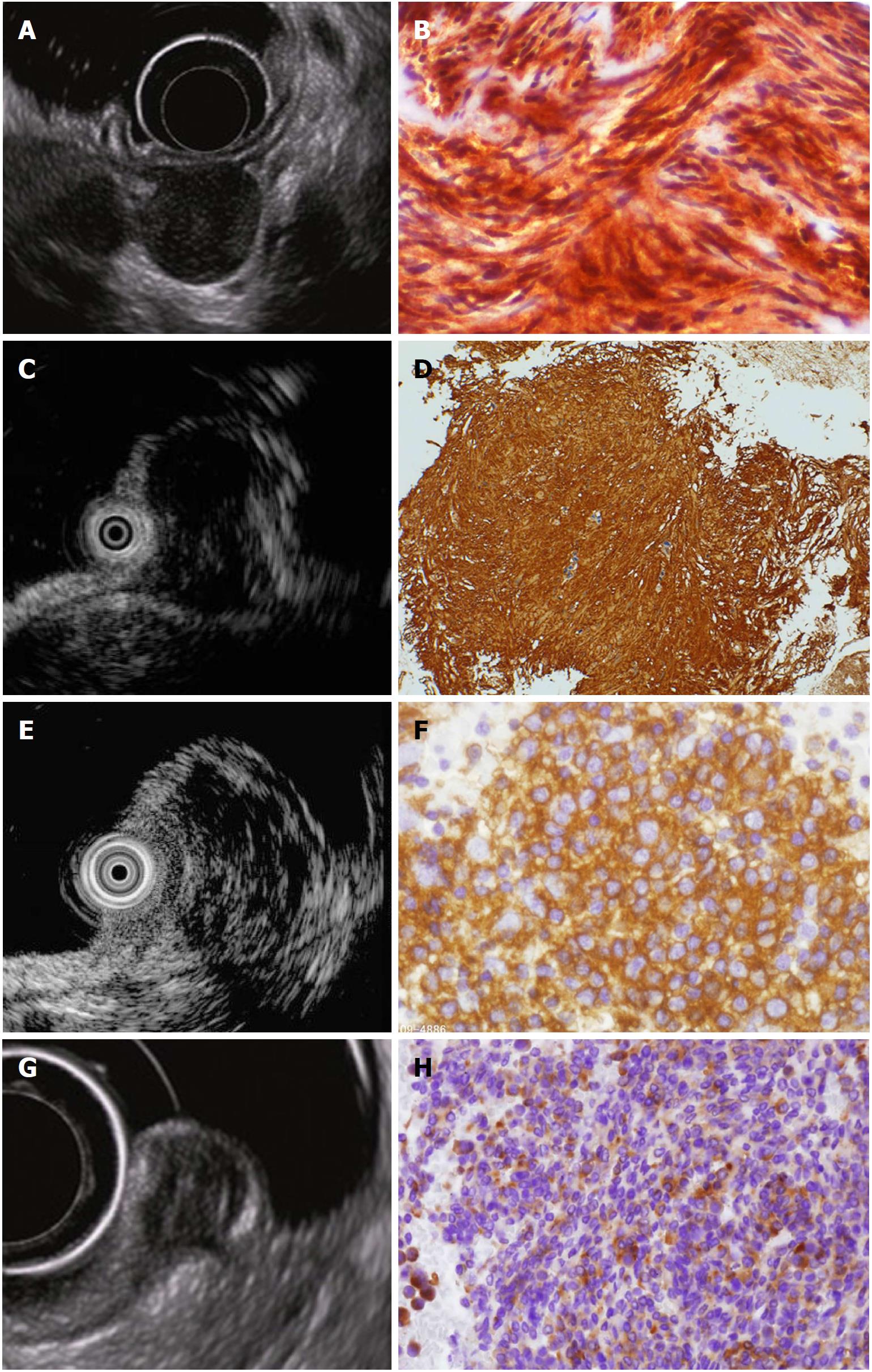
Figure 5 Endoscopic ultrasound images and corresponding endoscopic ultrasonography-guided fine needle aspiration specimens of hypoechoic solid tumors.
A: Endoscopic ultrasound (EUS) image of a gastric gastrointestinal stromal tumor; B: EUS-guided fine needle aspiration (EUS-FNA) specimen tissue image of A (KIT-positive spindle-shaped tumor cells are observed); C: EUS image of gastric leiomyoma; D: EUS-FNA specimen tissue image of C [α-SMA-positive spindle-shaped tumor cells are observed; diagnosis of leiomyoma was made by immunohistochemical analysis, which revealed α-SMA (+), KIT (-), CD34 (-), and S-100 (-)]; E: EUS image of gastric malignant lymphoma; F: EUS-FNA specimen image of E (diagnosis of diffuse large B-cell lymphoma was made by CD20-positive lymphoid tumor cells); G: EUS image of rectal neuroendocrine tumor (NET); H: EUS-FNA specimen image of G (diagnosis of NET was made by typical findings of irregular nest of synaptophysin-positive epithelial-like cells). Quoted and modified from reference[38] with permission.
- Citation: Akahoshi K, Oya M, Koga T, Shiratsuchi Y. Current clinical management of gastrointestinal stromal tumor. World J Gastroenterol 2018; 24(26): 2806-2817
- URL: https://www.wjgnet.com/1007-9327/full/v24/i26/2806.htm
- DOI: https://dx.doi.org/10.3748/wjg.v24.i26.2806


