Copyright
©The Author(s) 2018.
World J Gastroenterol. May 14, 2018; 24(18): 2009-2023
Published online May 14, 2018. doi: 10.3748/wjg.v24.i18.2009
Published online May 14, 2018. doi: 10.3748/wjg.v24.i18.2009
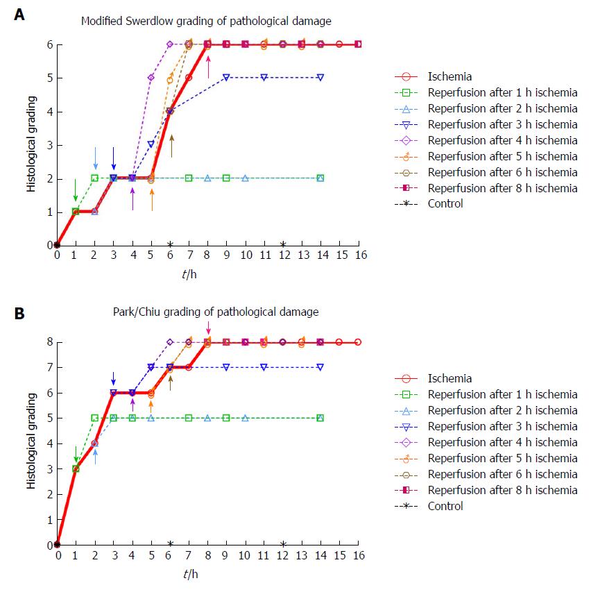
Figure 4 Histological grading of pathological damage (5 pigs, n = 128 biopsies total) at selected ischemia/reperfusion intervals.
Colored arrows show time points for start of reperfusion. Stippled lines show progression of injury following reperfusion. A: Modified Swerdlow et al[21,27,28]. B: Park/Chiu et al[22,26].
- Citation: Strand-Amundsen RJ, Reims HM, Reinholt FP, Ruud TE, Yang R, Høgetveit JO, Tønnessen TI. Ischemia/reperfusion injury in porcine intestine - Viability assessment. World J Gastroenterol 2018; 24(18): 2009-2023
- URL: https://www.wjgnet.com/1007-9327/full/v24/i18/2009.htm
- DOI: https://dx.doi.org/10.3748/wjg.v24.i18.2009


