Copyright
©The Author(s) 2017.
World J Gastroenterol. Nov 14, 2017; 23(42): 7609-7617
Published online Nov 14, 2017. doi: 10.3748/wjg.v23.i42.7609
Published online Nov 14, 2017. doi: 10.3748/wjg.v23.i42.7609
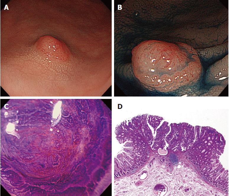
Figure 3 An early “missed or new” post-colonoscopy colorectal cancer case (No.
7 in Table 2). A 79-year-old man underwent initial colonoscopy, and seven small adenomatous polyps in the ascending and sigmoid colon were resected. A: A diminutive lesion 4 mm in size was found in the sigmoid colon during surveillance colonoscopy 15 mo after initial colonoscopy; B: Chromoendoscopy with indigo-carmine dye visualizes the margin of the deep depressed area on the surface of the lesion, and crystal violet stain shows a type-Vi pit with an invasive pattern suggesting submucosal deep invasive cancer (C); D: Histopathological examination of the surgical specimen reveals well to moderately differentiated adenocarcinoma with submucosal deep (3000 μm) invasion and no lymph node metastasis.
- Citation: Iwatate M, Kitagawa T, Katayama Y, Tokutomi N, Ban S, Hattori S, Hasuike N, Sano W, Sano Y, Tamano M. Post-colonoscopy colorectal cancer rate in the era of high-definition colonoscopy. World J Gastroenterol 2017; 23(42): 7609-7617
- URL: https://www.wjgnet.com/1007-9327/full/v23/i42/7609.htm
- DOI: https://dx.doi.org/10.3748/wjg.v23.i42.7609


