Copyright
©The Author(s) 2017.
World J Gastroenterol. Jul 7, 2017; 23(25): 4615-4623
Published online Jul 7, 2017. doi: 10.3748/wjg.v23.i25.4615
Published online Jul 7, 2017. doi: 10.3748/wjg.v23.i25.4615
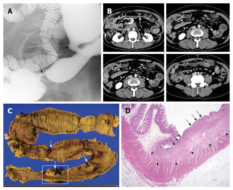
Figure 1 A 54-year-old man with abdominal pain and anemia.
A: Small bowel series spot compression image demonstrates multiple short strictures along the ileum (arrows); B: On axial images of contrast-enhanced computed tomography (CT) enterography, short-segmental strictures (arrows) show moderate and layered bowel wall enhancement with mild dilatation of intervening segment between strictures; C: Gross specimen of resected small intestine shows multiple short segmental strictures (arrows) with mild dilatation of intervening bowel segment, which corresponds with CT images (arrows in B); D: On the low-power H&E staining of surgical specimen (boxed area in Figure 1C) there is a superficial ulcer (arrows) and submucosal fibrosis (arrow heads) without evidence of transmural inflammation or granulomatous lesion (magnification × 10).
- Citation: Hwang J, Kim JS, Kim AY, Lim JS, Kim SH, Kim MJ, Kim MS, Song KD, Woo JY. Cryptogenic multifocal ulcerous stenosing enteritis: Radiologic features and clinical behavior. World J Gastroenterol 2017; 23(25): 4615-4623
- URL: https://www.wjgnet.com/1007-9327/full/v23/i25/4615.htm
- DOI: https://dx.doi.org/10.3748/wjg.v23.i25.4615


