Copyright
©The Author(s) 2016.
World J Gastroenterol. Nov 28, 2016; 22(44): 9752-9764
Published online Nov 28, 2016. doi: 10.3748/wjg.v22.i44.9752
Published online Nov 28, 2016. doi: 10.3748/wjg.v22.i44.9752
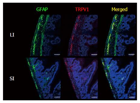
Figure 2 Enlarged view of GFAP/TRPV1-stained small intestine and large intestine of wild-type mice.
Certain parts of Figure 1 were magnified. GFAP is located in the myenteric plexus, submucosal plexus and fibrous structures penetrating into the circular muscle layer and mucosal layer (in WT LI, the serosal membrane was also stained with GFAP; however, serosal membrane is known to show false-positive immunosignals with various antibodies). The location of the TRPV1-IR signal coincided with some portions of the GFAP-IR signal in the myenteric plexus. Nuclei were stained with TO-PRO3. Representative data from 2 experiments using 2 mice per time point are shown. Scale bar represents 50 μm. GFAP: Glial fibrillary acidic protein; TRPV1: Transient receptor potential vanilloid 1; SI: Small intestine; LI: Large intestine; WT: Wild-type.
- Citation: Yamamoto M, Nishiyama M, Iizuka S, Suzuki S, Suzuki N, Aiso S, Nakahara J. Transient receptor potential vanilloid 1-immunoreactive signals in murine enteric glial cells. World J Gastroenterol 2016; 22(44): 9752-9764
- URL: https://www.wjgnet.com/1007-9327/full/v22/i44/9752.htm
- DOI: https://dx.doi.org/10.3748/wjg.v22.i44.9752


