Copyright
©The Author(s) 2016.
World J Gastroenterol. Nov 7, 2016; 22(41): 9127-9140
Published online Nov 7, 2016. doi: 10.3748/wjg.v22.i41.9127
Published online Nov 7, 2016. doi: 10.3748/wjg.v22.i41.9127
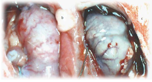
Figure 9 Blood vessels presentation at the ventral site of the stomach surface in rats that just underwent esophagogastric anastomosis.
In general, immediately after anastomosis creation and saline bath application, blood vessels disappeared from the rat gastric surface, and this effect lasts at least the next 15 min (scored Min/Med/Max 0/0/0), a period that was carefully monitored (control, c). With a BPC 157 bath immediately after anastomosis creation, the blood vessels did not disappear from the rat gastric surface; instead, these vessels remained present during at least the next 15 min of monitoring (BPC 157, b).
- Citation: Djakovic Z, Djakovic I, Cesarec V, Madzarac G, Becejac T, Zukanovic G, Drmic D, Batelja L, Zenko Sever A, Kolenc D, Pajtak A, Knez N, Japjec M, Luetic K, Stancic-Rokotov D, Seiwerth S, Sikiric P. Esophagogastric anastomosis in rats: Improved healing by BPC 157 and L-arginine, aggravated by L-NAME. World J Gastroenterol 2016; 22(41): 9127-9140
- URL: https://www.wjgnet.com/1007-9327/full/v22/i41/9127.htm
- DOI: https://dx.doi.org/10.3748/wjg.v22.i41.9127


