Copyright
©The Author(s) 2016.
World J Gastroenterol. Jan 28, 2016; 22(4): 1701-1710
Published online Jan 28, 2016. doi: 10.3748/wjg.v22.i4.1701
Published online Jan 28, 2016. doi: 10.3748/wjg.v22.i4.1701
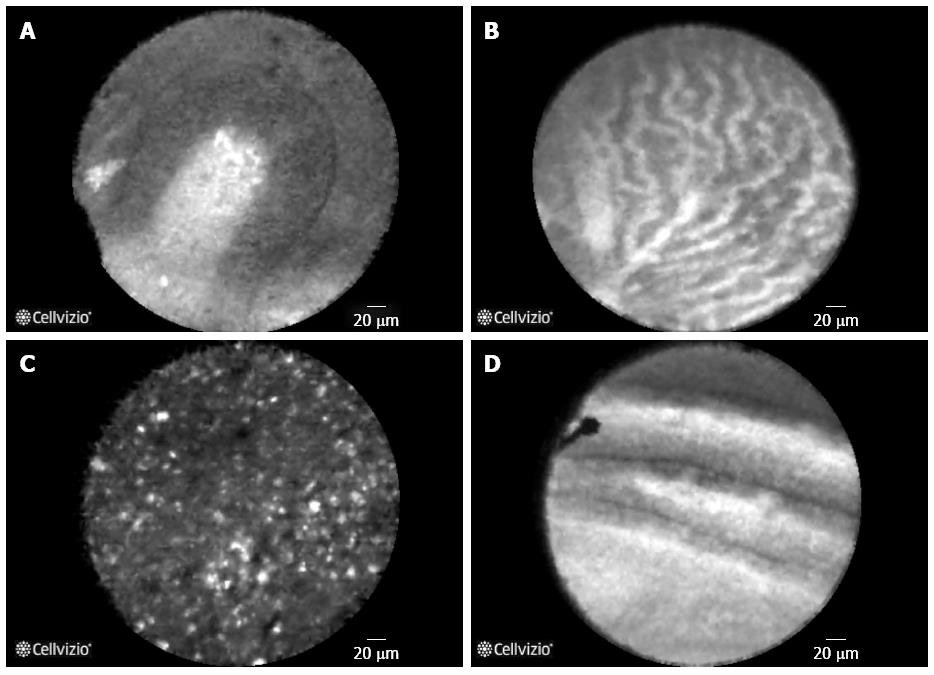
Figure 1 Different types of pancreatic cysts.
A: Intraductal papillary mucinous neoplasm. A single papilla is visualized with a central fibrovascular core and overlying epithelium; B: Serous cystadenoma: Branching and tortuous network of multiple blood vessels in a “fern like” pattern. This is has been termed as “superficial vascular network”[22]; C: Pseudocyst: Clusters of bright, floating particles with a background which is nondescript and lacks blood vessels; D: Mucinous cystic neoplasm: Solitary epithelial bands without formation of papillae.
- Citation: Krishna SG, Lee JH. Appraisal of needle-based confocal laser endomicroscopy in the diagnosis of pancreatic cysts. World J Gastroenterol 2016; 22(4): 1701-1710
- URL: https://www.wjgnet.com/1007-9327/full/v22/i4/1701.htm
- DOI: https://dx.doi.org/10.3748/wjg.v22.i4.1701


