Copyright
©The Author(s) 2016.
World J Gastroenterol. Sep 7, 2016; 22(33): 7507-7517
Published online Sep 7, 2016. doi: 10.3748/wjg.v22.i33.7507
Published online Sep 7, 2016. doi: 10.3748/wjg.v22.i33.7507
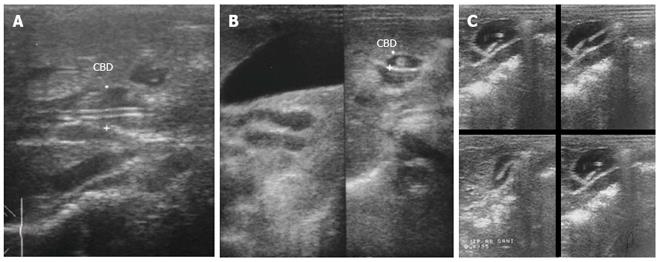
Figure 12 Ultrasonography images in hepatobiliary and pancreatic ascariasis.
A: Biliary ultrasound depicting four-line sign; B: Ultrasound showing tube like structure within common bile duct with distended gall bladder; C: Ascarides in gall bladder showing active movements as seen in serial images. Adapted from Khuroo[5].
- Citation: Khuroo MS, Rather AA, Khuroo NS, Khuroo MS. Hepatobiliary and pancreatic ascariasis. World J Gastroenterol 2016; 22(33): 7507-7517
- URL: https://www.wjgnet.com/1007-9327/full/v22/i33/7507.htm
- DOI: https://dx.doi.org/10.3748/wjg.v22.i33.7507


