Copyright
©The Author(s) 2016.
World J Gastroenterol. Jun 28, 2016; 22(24): 5479-5494
Published online Jun 28, 2016. doi: 10.3748/wjg.v22.i24.5479
Published online Jun 28, 2016. doi: 10.3748/wjg.v22.i24.5479
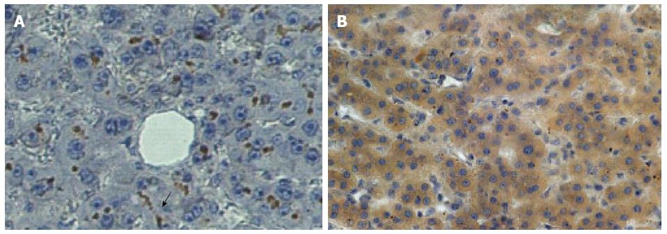
Figure 5 Distribution of CEACAM1 (BGP) in human liver.
Photomicrographs of CEACAM1 staining with monoclonal antibody 4D1/C2 showing very intense brown staining mainly in the distribution of the biliary canaliculus in normal human liver (A).In contrast, in the normal portion of a liver from a patient with hepatic metastasis (B), dark staining is seen in the cytoplasm of the hepatocytes with no canalicular staining evident. Published with permission of Springer[8]. The arrow points to the typical distribution of bound ant-BGP in the bile canaliculus in the normal liver (A) at center, bottom.
- Citation: Tobi M, Thomas P, Ezekwudo D. Avoiding hepatic metastasis naturally: Lessons from the cotton top tamarin (Saguinus oedipus). World J Gastroenterol 2016; 22(24): 5479-5494
- URL: https://www.wjgnet.com/1007-9327/full/v22/i24/5479.htm
- DOI: https://dx.doi.org/10.3748/wjg.v22.i24.5479


