Copyright
©The Author(s) 2015.
World J Gastroenterol. Oct 7, 2015; 21(37): 10553-10562
Published online Oct 7, 2015. doi: 10.3748/wjg.v21.i37.10553
Published online Oct 7, 2015. doi: 10.3748/wjg.v21.i37.10553
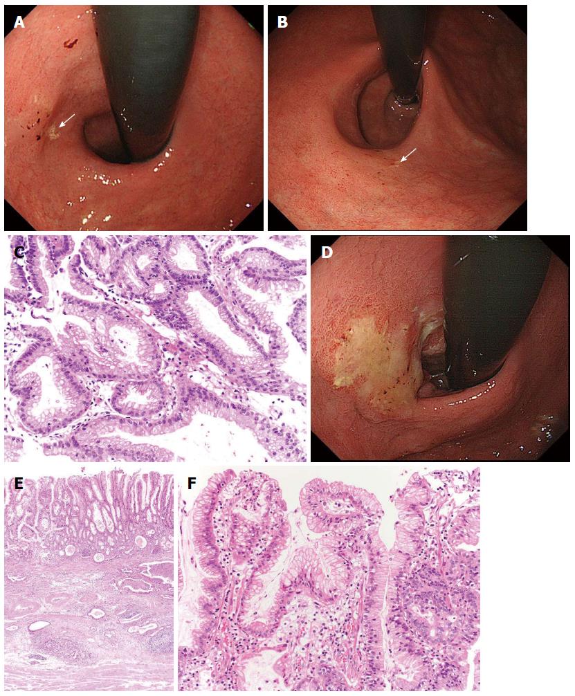
Figure 5 Gastric cancer progressed after eradication therapy.
A: A review of the endoscopic pictures taken 3 years earlier revealed the presence of a discolored and flat elevated lesion adjacent to a xanthoma (arrow) in the cardia; B: This lesion was not clear yet, but the xanthoma had almost disappeared (arrow); C: The biopsy specimen taken from the lesion showed foveolar-type epithelium with mild structural atypia; D: After follow-up for 1 yr, the tumor had become evident and covered by mucus; E: The tumor had invaded into the deep submucosal layer, but the mucosal element was preserved; F: This cancer showed a gastric mucin phenotype with low-grade atypia[26].
-
Citation: Kobayashi M, Sato Y, Terai S. Endoscopic surveillance of gastric cancers after
Helicobacter pylori eradication. World J Gastroenterol 2015; 21(37): 10553-10562 - URL: https://www.wjgnet.com/1007-9327/full/v21/i37/10553.htm
- DOI: https://dx.doi.org/10.3748/wjg.v21.i37.10553


