Copyright
©The Author(s) 2015.
World J Gastroenterol. Oct 7, 2015; 21(37): 10553-10562
Published online Oct 7, 2015. doi: 10.3748/wjg.v21.i37.10553
Published online Oct 7, 2015. doi: 10.3748/wjg.v21.i37.10553
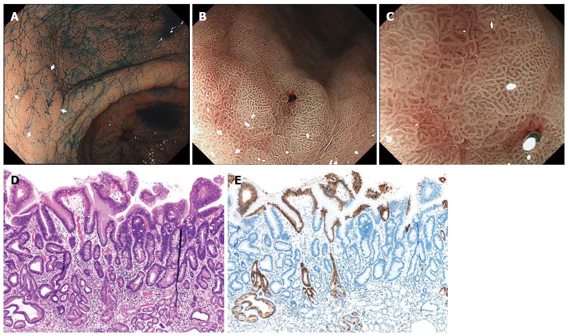
Figure 4 Differentiated-type early gastric cancer detected after successful Helicobacter pylori eradication.
A: Chromoendoscopy revealed a depressed-type lesion in the anterior wall of the antrum; B, C: Narrow-band imaging with magnified endoscopy shows a “gastritis-like” appearance with a microsurface structure comprising of regular papillae resembling with unclear demarcation between the cancer and surrounding gastritis mucosa; D: Well-differentiated tubular adenocarcinoma with low-grade atypia (hematoxylin and eosin staining); E: MUC5AC immunohistochemical staining demonstrates that superficial non-neoplastic epithelium is interspersed among and above the cancer tubules[47].
-
Citation: Kobayashi M, Sato Y, Terai S. Endoscopic surveillance of gastric cancers after
Helicobacter pylori eradication. World J Gastroenterol 2015; 21(37): 10553-10562 - URL: https://www.wjgnet.com/1007-9327/full/v21/i37/10553.htm
- DOI: https://dx.doi.org/10.3748/wjg.v21.i37.10553


