Copyright
©The Author(s) 2015.
World J Gastroenterol. Oct 7, 2015; 21(37): 10553-10562
Published online Oct 7, 2015. doi: 10.3748/wjg.v21.i37.10553
Published online Oct 7, 2015. doi: 10.3748/wjg.v21.i37.10553
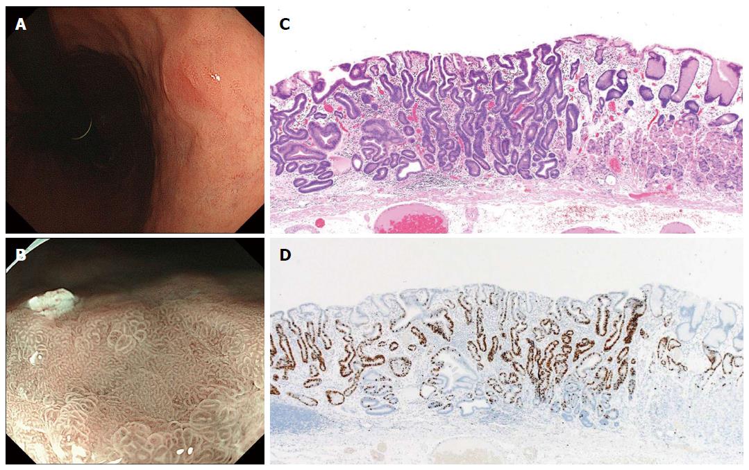
Figure 3 Differentiated-type early gastric cancer detected after successful Helicobacter pylori eradication.
A: This endoscopic finding made qualitative cancer diagnosis difficult by conventional endoscopy; B: A “gastritis-like” appearance under narrow-band imaging with magnified endoscopy was demonstrated by a microsurface structure comprised of mixed pits and papillae, resembling the surrounding non-cancerous mucosa; C: Well-differentiated tubular adenocarcinoma with low-grade atypia (hematoxylin and eosin staining); D: Ki-67 positive cells localized in the middle-to-lower portion of the mucosa, indicating cytological maturation at the luminal surface layer of the cancer[47].
-
Citation: Kobayashi M, Sato Y, Terai S. Endoscopic surveillance of gastric cancers after
Helicobacter pylori eradication. World J Gastroenterol 2015; 21(37): 10553-10562 - URL: https://www.wjgnet.com/1007-9327/full/v21/i37/10553.htm
- DOI: https://dx.doi.org/10.3748/wjg.v21.i37.10553


