Copyright
©The Author(s) 2015.
World J Gastroenterol. Aug 28, 2015; 21(32): 9512-9525
Published online Aug 28, 2015. doi: 10.3748/wjg.v21.i32.9512
Published online Aug 28, 2015. doi: 10.3748/wjg.v21.i32.9512
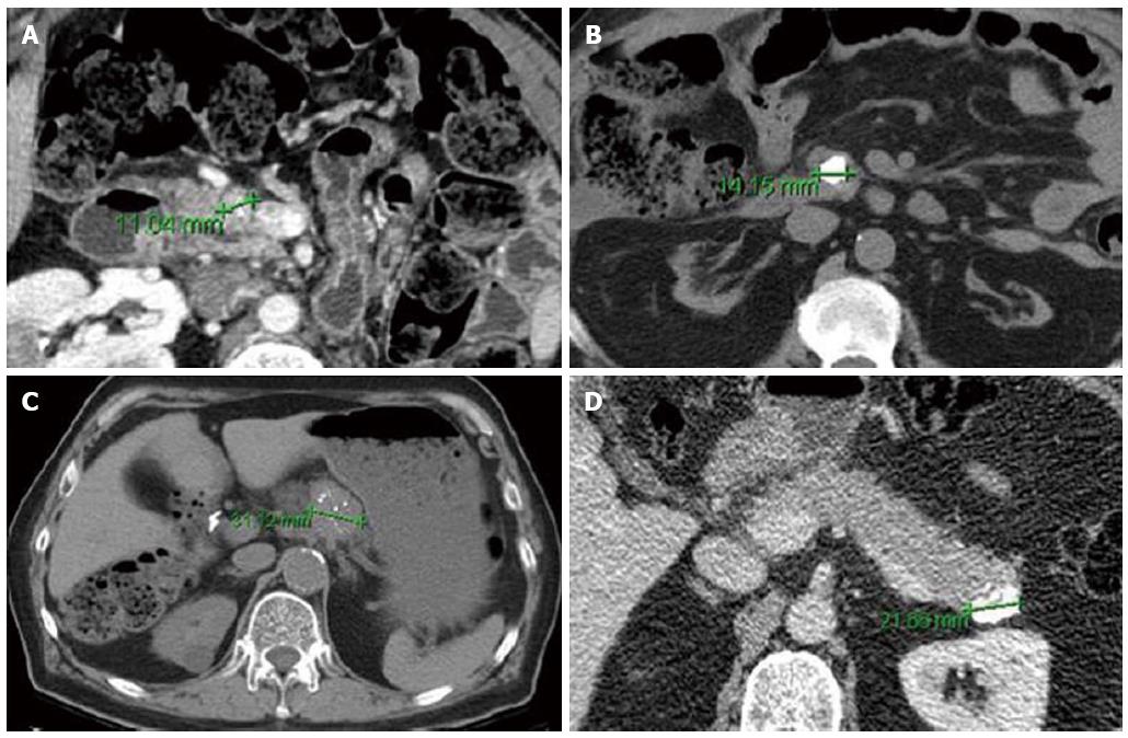
Figure 2 Axial computed tomography images of Pancreatic neuroendocrine tumor with punctate (A, C) and dense/coarse calcifications (B, D).
Despite their small size, all lesions were associated with either lymph node metastasis (A-C) or intermediate (G2) grade (B-D) on pathologic evaluation (From Poultsides et al[36]. Pancreatic Neuroendocrine Tumors: Radiographic Calcifications Correlate with Grade and Metastasis. Ann Surg Onc 2012; 19: 2295-2303).
- Citation: Cloyd JM, Poultsides GA. Non-functional neuroendocrine tumors of the pancreas: Advances in diagnosis and management. World J Gastroenterol 2015; 21(32): 9512-9525
- URL: https://www.wjgnet.com/1007-9327/full/v21/i32/9512.htm
- DOI: https://dx.doi.org/10.3748/wjg.v21.i32.9512


