Copyright
©The Author(s) 2015.
World J Gastroenterol. Aug 28, 2015; 21(32): 9512-9525
Published online Aug 28, 2015. doi: 10.3748/wjg.v21.i32.9512
Published online Aug 28, 2015. doi: 10.3748/wjg.v21.i32.9512
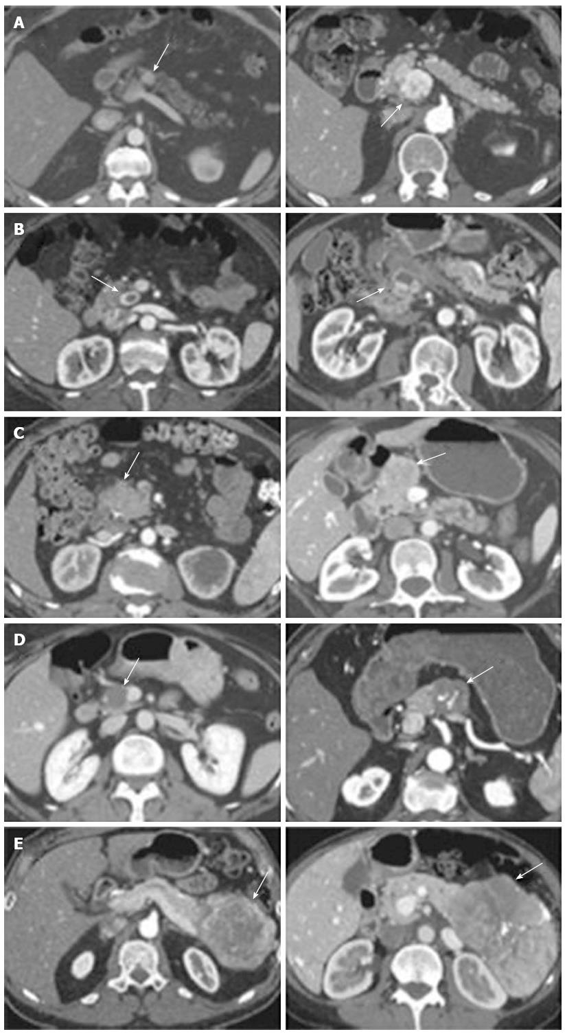
Figure 1 Representative images of the 5 types of pancreatic neuroendocrine tumor enhancement pattern on arterial phase computed tomography.
Two images are shown for each type. A: Hyperenhancing, solid; B: Cystic with hyperenhancing rim; C: Isoenhancing or no mass visualized; D: Homogeneously hypoenhancing; E: Heterogeneous but mostly hypoenhancing with some peripheral enhancement. Groups D and E had worse survival after resection compared with groups A, B, and C (From Worhunsky et al[35]. Pancreatic neuroendocrine tumours: hypoenhancement on arterial phase computed tomography predicts biological aggressiveness. HPB 2014; 16: 304-311). Arrows indicate PNET.
- Citation: Cloyd JM, Poultsides GA. Non-functional neuroendocrine tumors of the pancreas: Advances in diagnosis and management. World J Gastroenterol 2015; 21(32): 9512-9525
- URL: https://www.wjgnet.com/1007-9327/full/v21/i32/9512.htm
- DOI: https://dx.doi.org/10.3748/wjg.v21.i32.9512


