Copyright
©The Author(s) 2015.
World J Gastroenterol. Jan 14, 2015; 21(2): 675-687
Published online Jan 14, 2015. doi: 10.3748/wjg.v21.i2.675
Published online Jan 14, 2015. doi: 10.3748/wjg.v21.i2.675
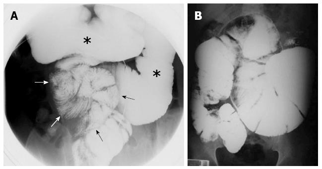
Figure 3 Small bowel transit.
Procubitus image with localized compression. Liquid distension of the gastroduodenum (asterisks) and adhesion of the small intestinal loops (arrows) are persistent despite localized compression, producing a “cauliflower” appearance[24]; B: Upper gastrointestinal images reveal dilatation of the duodenum and jejunal loops, delayed bowel transit, and failure of the oral contrast to pass distally[38].
- Citation: Akbulut S. Accurate definition and management of idiopathic sclerosing encapsulating peritonitis. World J Gastroenterol 2015; 21(2): 675-687
- URL: https://www.wjgnet.com/1007-9327/full/v21/i2/675.htm
- DOI: https://dx.doi.org/10.3748/wjg.v21.i2.675


