Copyright
©2014 Baishideng Publishing Group Inc.
World J Gastroenterol. Dec 28, 2014; 20(48): 18216-18227
Published online Dec 28, 2014. doi: 10.3748/wjg.v20.i48.18216
Published online Dec 28, 2014. doi: 10.3748/wjg.v20.i48.18216
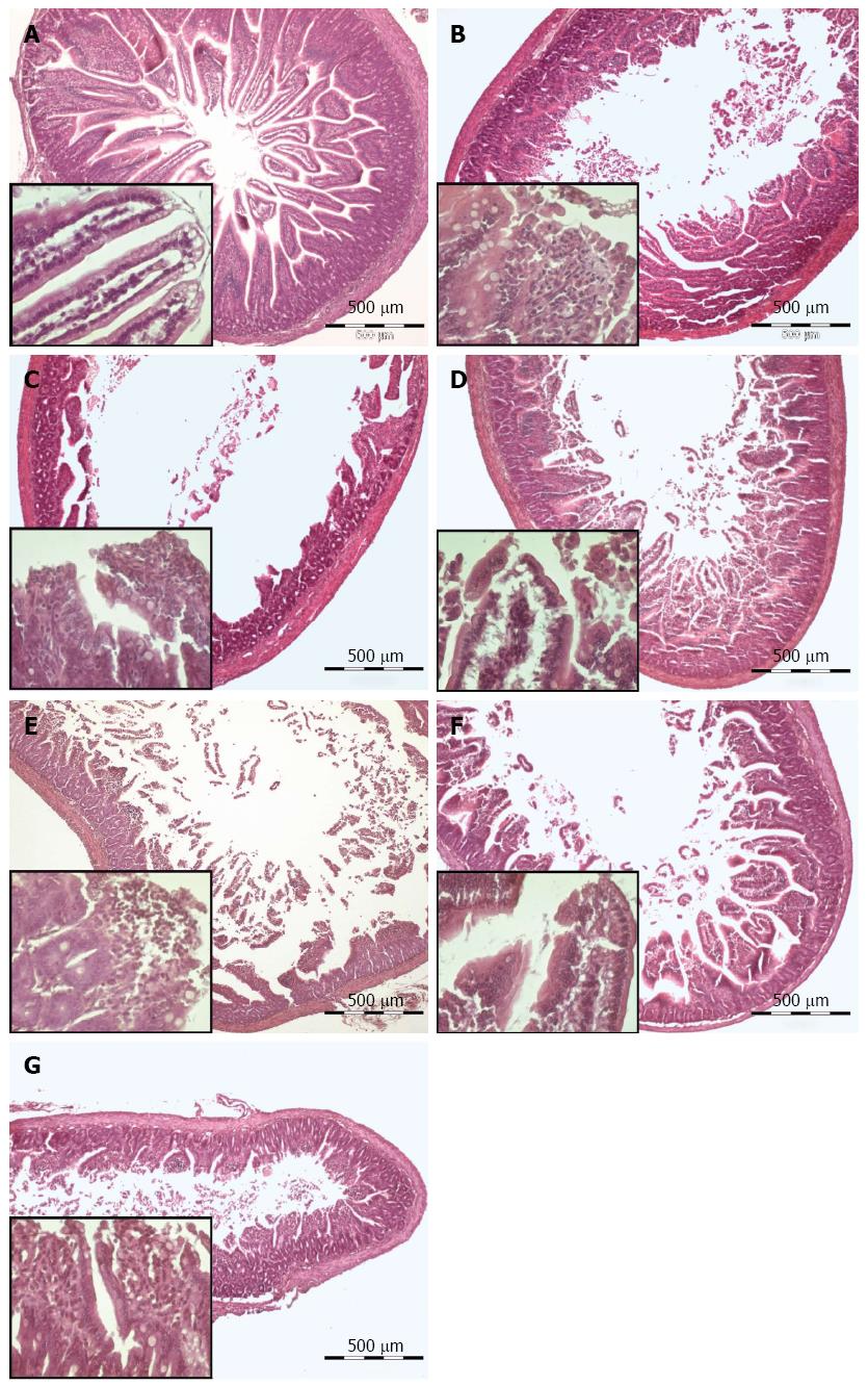
Figure 8 Effect of AQIX, MEM-HEPES and Tyrode solution (Hematoxylin-Eosin staining).
A, B, D, F after storage in the respective solution; C, E,G a cuff of the same segment after perfusion with the same solution. In the lower left quarter of each picture the mucosal structure is shown in detail. A: Unperfused tissue; B: Stored in Tyrode solution; C: Stored and perfused with Tyrode solution; D: Stored in MEM-Hepes medium; E: Stored and perfused with MEM-Hepes medium; F: Stored in AQIX RS-I solution; G: Stored and perfused with AQIX RS-I solution.
-
Citation: Schreiber D, Jost V, Bischof M, Seebach K, Lammers WJ, Douglas R, Schäfer KH. Motility patterns of
ex vivo intestine segments depend on perfusion mode. World J Gastroenterol 2014; 20(48): 18216-18227 - URL: https://www.wjgnet.com/1007-9327/full/v20/i48/18216.htm
- DOI: https://dx.doi.org/10.3748/wjg.v20.i48.18216


