Copyright
©2014 Baishideng Publishing Group Inc.
World J Gastroenterol. Oct 14, 2014; 20(38): 13842-13862
Published online Oct 14, 2014. doi: 10.3748/wjg.v20.i38.13842
Published online Oct 14, 2014. doi: 10.3748/wjg.v20.i38.13842
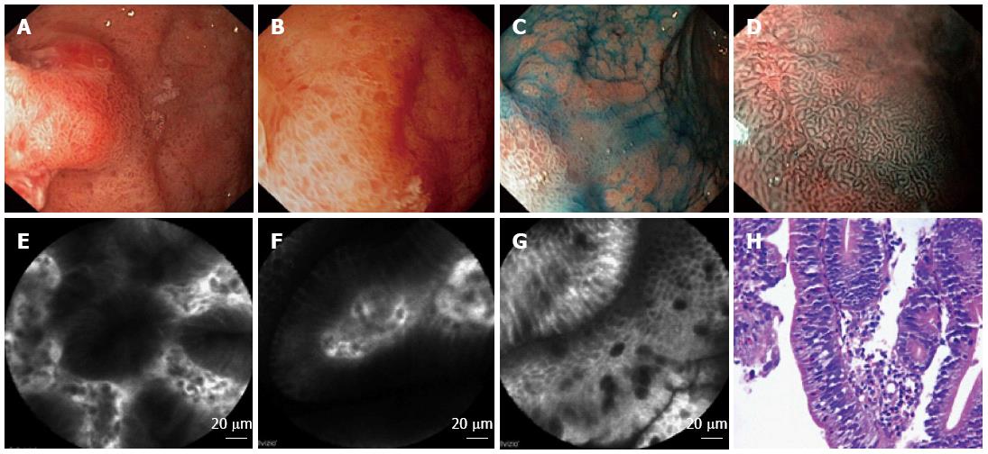
Figure 3 Characterization of the surrounding mucosa of the same patient.
A-C: WLE + chromoendoscopy: pronounced focal mucosal hyperemia, edema, petechiae, multiply foci of intestinal metaplasia; D: Narrow band imaging (NBI) + magnified endoscopy (zoom). The results, according to VS-classification[138]: the surrounding mucosa is inflamed, with a regular stick-like microsurface pattern, slightly irregular wavy microvascular pattern; E: Confocal laser endomicroscopy (CLE): A cross-section of normal glands; F: CLE: A longitudinal section of normal glands; G: CLE: Marks of intestinal metaplasia - Goblet cells; H: Pathomorphology: Active chronic Нр+ gastritis with incomplete intestinal metaplasia and low grade epithelial dysplasia.
- Citation: Pasechnikov V, Chukov S, Fedorov E, Kikuste I, Leja M. Gastric cancer: Prevention, screening and early diagnosis. World J Gastroenterol 2014; 20(38): 13842-13862
- URL: https://www.wjgnet.com/1007-9327/full/v20/i38/13842.htm
- DOI: https://dx.doi.org/10.3748/wjg.v20.i38.13842


