Copyright
©2014 Baishideng Publishing Group Co.
World J Gastroenterol. May 14, 2014; 20(18): 5263-5273
Published online May 14, 2014. doi: 10.3748/wjg.v20.i18.5263
Published online May 14, 2014. doi: 10.3748/wjg.v20.i18.5263
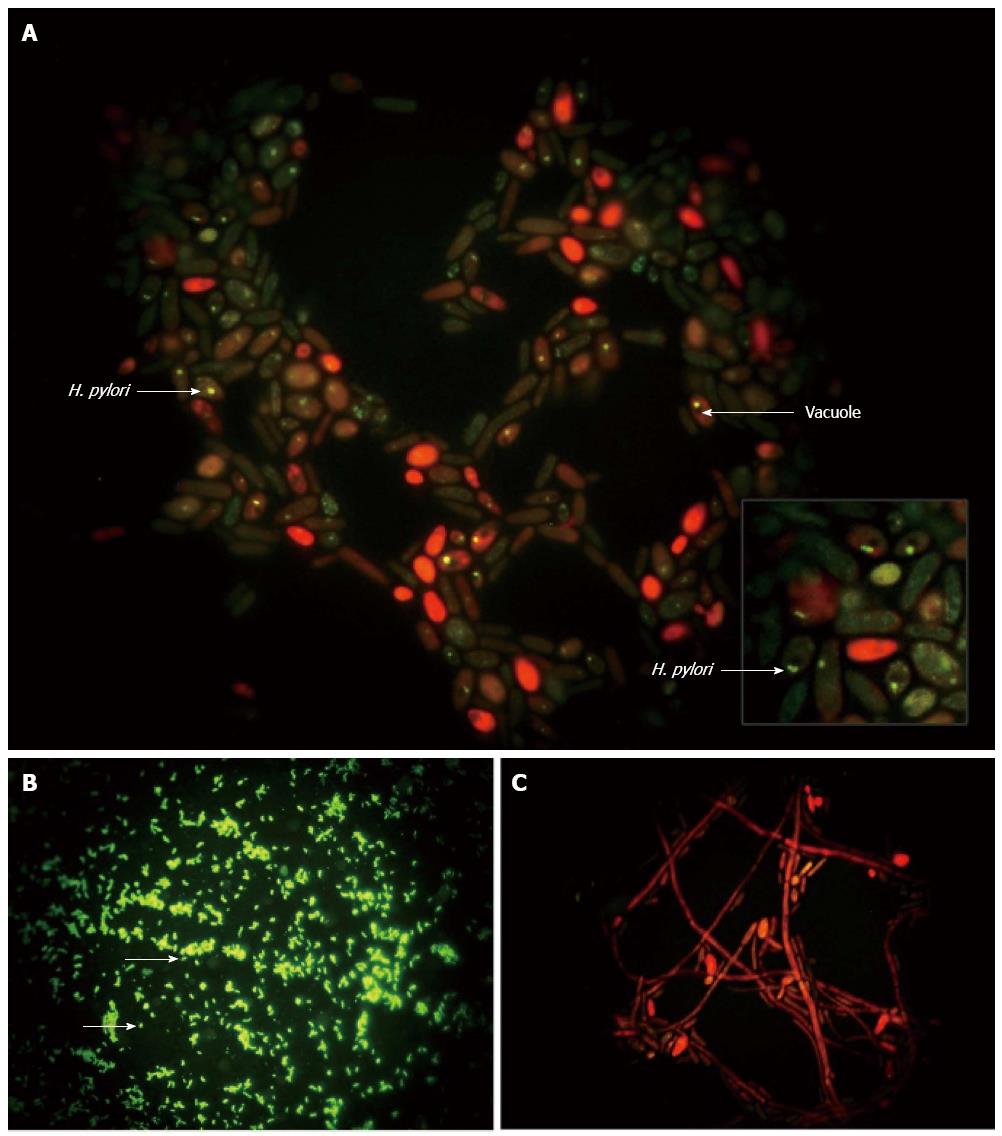
Figure 4 Immunolabeling of Helicobacter pylori.
Immunofluorescence micrographs showing: A: Localization of Helicobacter pylori (H. pylori) (green) inside the Candida yeast, some of which appear red due to diffusion of the counterstain (magnification × 4000); B: Control H. pylori (green; arrows) (magnification × 1000); and C: Absence of bacteria in heat-killed yeast (magnification × 1000)[108].
-
Citation: Siavoshi F, Saniee P. Vacuoles of
Candida yeast as a specialized niche forHelicobacter pylori . World J Gastroenterol 2014; 20(18): 5263-5273 - URL: https://www.wjgnet.com/1007-9327/full/v20/i18/5263.htm
- DOI: https://dx.doi.org/10.3748/wjg.v20.i18.5263


