Copyright
©2014 Baishideng Publishing Group Co.
World J Gastroenterol. May 14, 2014; 20(18): 5263-5273
Published online May 14, 2014. doi: 10.3748/wjg.v20.i18.5263
Published online May 14, 2014. doi: 10.3748/wjg.v20.i18.5263
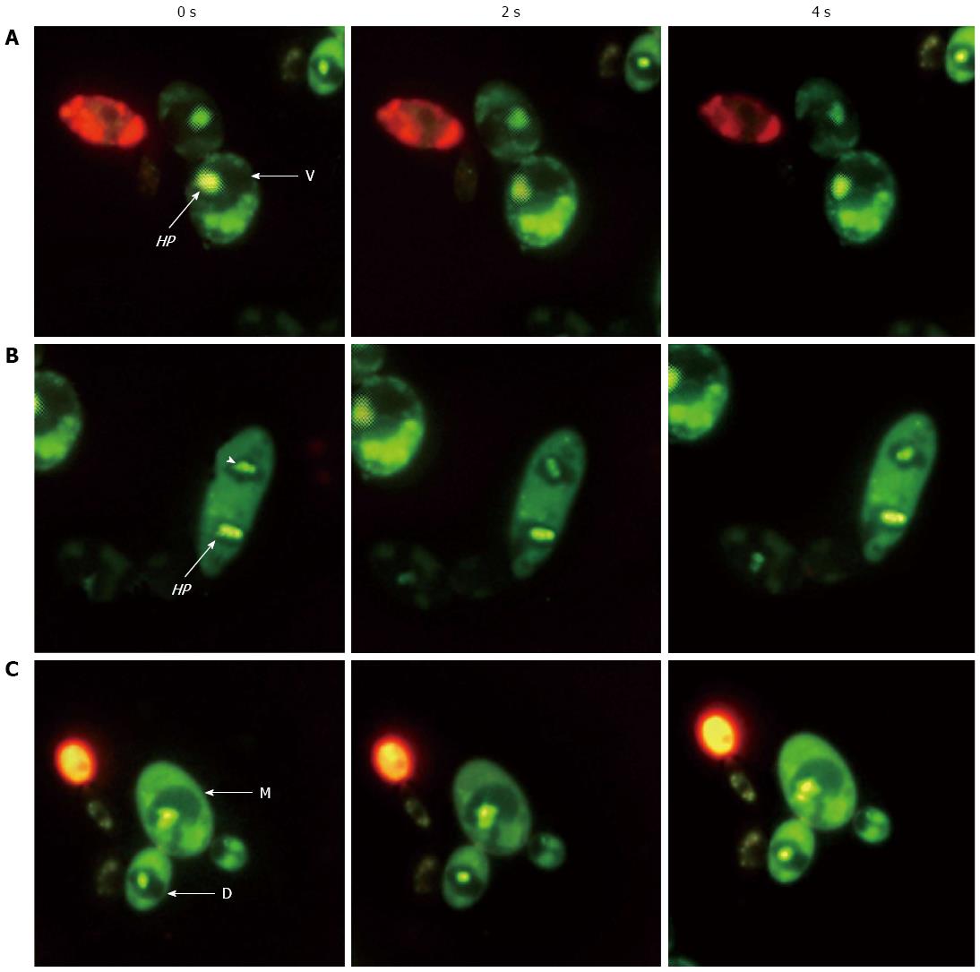
Figure 3 Helicobacter pylori movement in dividing yeast.
Fluorescence micrographs from three experiments (A, B and C) using a live/dead-BacLight kit showing live (green) fast-moving Helicobacter pylori (HP) cells (arrows and arrowhead) inside the vacuoles (V) of mother (M) and daughter (D) Candida cells taken at 0, 2 and 4 s (magnification × 1000)[108].
-
Citation: Siavoshi F, Saniee P. Vacuoles of
Candida yeast as a specialized niche forHelicobacter pylori . World J Gastroenterol 2014; 20(18): 5263-5273 - URL: https://www.wjgnet.com/1007-9327/full/v20/i18/5263.htm
- DOI: https://dx.doi.org/10.3748/wjg.v20.i18.5263


