Copyright
©2014 Baishideng Publishing Group Co.
World J Gastroenterol. Apr 28, 2014; 20(16): 4467-4482
Published online Apr 28, 2014. doi: 10.3748/wjg.v20.i16.4467
Published online Apr 28, 2014. doi: 10.3748/wjg.v20.i16.4467
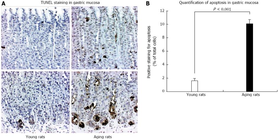
Figure 9 TUNEL staining for apoptosis in gastric mucosa of young and aging rats.
A: The photomicrographs of gastric mucosa of young and aging rats at baseline (magnification x 100). In situ cell death (apoptosis) detection by terminal deoxynucleotidyl transferase-mediated dUTP nick end labeling (TUNEL) was used to visualize apoptotic-positive cells (brown staining); B: Quantification of the number of positively labeled cells demonstrated that gastric mucosa of aging rats exhibits a significantly increased number of apoptotic cells vs mucosa of young rats. The increased apoptosis prominently involved epithelial cells at the basal mucosa explaining atrophy of the basal gastric glands shown in Figure 4. Reproduced with permission from Tarnawski et al[1].
- Citation: Tarnawski AS, Ahluwalia A, Jones MK. Increased susceptibility of aging gastric mucosa to injury: The mechanisms and clinical implications. World J Gastroenterol 2014; 20(16): 4467-4482
- URL: https://www.wjgnet.com/1007-9327/full/v20/i16/4467.htm
- DOI: https://dx.doi.org/10.3748/wjg.v20.i16.4467


