Copyright
©2014 Baishideng Publishing Group Co.
World J Gastroenterol. Apr 28, 2014; 20(16): 4467-4482
Published online Apr 28, 2014. doi: 10.3748/wjg.v20.i16.4467
Published online Apr 28, 2014. doi: 10.3748/wjg.v20.i16.4467
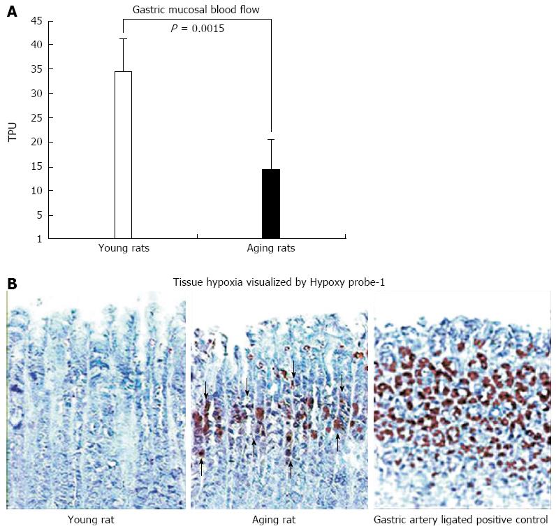
Figure 6 Gastric mucosal blood flow measured with BLF21 Laser-Doppler flow meter and mucosal hypoxia visualized by Hypoxy probe-1.
A: In gastric mucosa of aging rats at baseline, mucosal blood flow, expressed in perfusion units, is significantly reduced by approximation 60% (vs young rats; P = 0.0015). Such a dramatic reduction in blood flow likely leads to chronic hypoxia; B: Photomicrographs of rat gastric mucosa. Gastric mucosal hypoxia is visualized by immunohistochemical staining utilizing the small molecular marker, pimonidazole HCl (Hypoxy probe-1), which binds selectively to oxygen starved cells[30]. In young rats, Hypoxy probe-1 staining is negative in both connective tissue and epithelial cells of the gastric mucosa demonstrating the absence of hypoxia. In aging rats, positive staining is strongly expressed (brown staining) in the upper and mid-mucosa, mainly in the progenitor and parietal cell zone (arrows), reflecting severe hypoxia in these cells. As a positive control we used gastric mucosa of young rats that had all major gastric arteries ligated for 1 h. A strong accumulation Hypoxy probe-1 is present in the majority of epithelial cells (brown staining) reflecting profound cell hypoxia. Reproduced with permission from Tarnawski et al[1].
- Citation: Tarnawski AS, Ahluwalia A, Jones MK. Increased susceptibility of aging gastric mucosa to injury: The mechanisms and clinical implications. World J Gastroenterol 2014; 20(16): 4467-4482
- URL: https://www.wjgnet.com/1007-9327/full/v20/i16/4467.htm
- DOI: https://dx.doi.org/10.3748/wjg.v20.i16.4467


