Copyright
©2014 Baishideng Publishing Group Co.
World J Gastroenterol. Apr 28, 2014; 20(16): 4467-4482
Published online Apr 28, 2014. doi: 10.3748/wjg.v20.i16.4467
Published online Apr 28, 2014. doi: 10.3748/wjg.v20.i16.4467
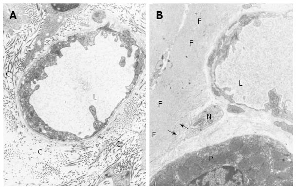
Figure 5 Transmission electron microscopy.
A: Transmission electron micrograph of perivascular connective tissue from a 3-month-old control rat from the basal portion of the oxyntic mucosa. The connective tissue shows numerous collagen fibers (C); L, Microvessel lumen. Magnification x 19000; B: Transmission electron micrograph of perivascular connective tissue from an old rat. In the basal portion of the oxyntic mucosa, collagen fibers are mostly absent and replaced by rudimentary collagen fibers (arrows) and deposits of amorphous fibrillar material (F). P: Parietal cells; L: Blood microvessel lumen; N: Nerve bundle. Magnification x 19000. Reproduced with permission from Hollander, Tarnawski et al[2].
- Citation: Tarnawski AS, Ahluwalia A, Jones MK. Increased susceptibility of aging gastric mucosa to injury: The mechanisms and clinical implications. World J Gastroenterol 2014; 20(16): 4467-4482
- URL: https://www.wjgnet.com/1007-9327/full/v20/i16/4467.htm
- DOI: https://dx.doi.org/10.3748/wjg.v20.i16.4467


