Copyright
©2014 Baishideng Publishing Group Co.
World J Gastroenterol. Apr 28, 2014; 20(16): 4467-4482
Published online Apr 28, 2014. doi: 10.3748/wjg.v20.i16.4467
Published online Apr 28, 2014. doi: 10.3748/wjg.v20.i16.4467
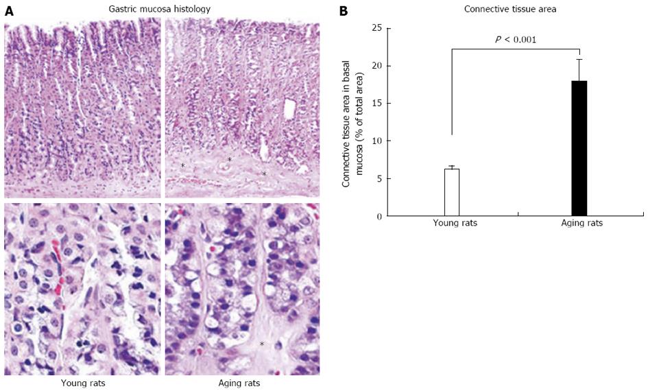
Figure 4 Photomicrographs of gastric mucosa in young and aging rats.
In gastric mucosa of aging rats there is partial atrophy of gastric glands in the basal mucosa and their replacement with connective tissue (*). A: Hematoxylin and eosin staining at low magnification (x 100) is shown in the upper panels and higher magnification (x 500) is shown in the lower panels; B: Quantification of connective tissue in the lower one third of the gastric mucosa shows a significant increase in connective tissue replacing glandular cells in aging rats. Quantification of the number of inflammatory cells in gastric mucosa shows no inflammation (only a minimal number of inflammatory cells) and no difference between young and aging rats indicating that atrophic changes are not accompanied by an inflammation. Reproduced with permission from Tarnawski et al[1].
- Citation: Tarnawski AS, Ahluwalia A, Jones MK. Increased susceptibility of aging gastric mucosa to injury: The mechanisms and clinical implications. World J Gastroenterol 2014; 20(16): 4467-4482
- URL: https://www.wjgnet.com/1007-9327/full/v20/i16/4467.htm
- DOI: https://dx.doi.org/10.3748/wjg.v20.i16.4467


