Copyright
©2014 Baishideng Publishing Group Co.
World J Gastroenterol. Apr 21, 2014; 20(15): 4300-4315
Published online Apr 21, 2014. doi: 10.3748/wjg.v20.i15.4300
Published online Apr 21, 2014. doi: 10.3748/wjg.v20.i15.4300
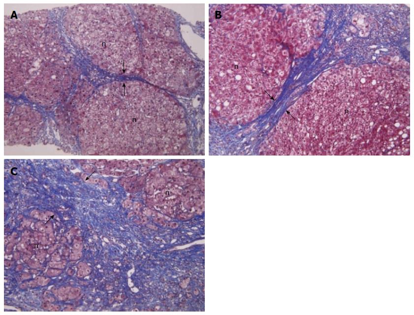
Figure 1 Heterogeneity of cirrhosis in histology.
All pictures are histological finding of cirrhosis however, they show different feature in thickness of sepata and the size of nodules. A: Shows mild cirrhosis with thin septa (MTC stain, × 100); B: Shows moderate cirrhosis with at least two broad septa; C: Shows severe cirrhosis with at least one very broad septa (MTC stain, respectively, × 200). A, B, C are correspond to subclass of Laennec fibrosis scoring system F4A, F4B and F4C respectively, The widths between two arrows show the significant difference among subclass of cirrhosis. n: Regenerating nodule; MTC: Masson trichrome stain[8].
- Citation: Kim MY, Jeong WK, Baik SK. Invasive and non-invasive diagnosis of cirrhosis and portal hypertension. World J Gastroenterol 2014; 20(15): 4300-4315
- URL: https://www.wjgnet.com/1007-9327/full/v20/i15/4300.htm
- DOI: https://dx.doi.org/10.3748/wjg.v20.i15.4300


