Copyright
©2014 Baishideng Publishing Group Co.
World J Gastroenterol. Apr 7, 2014; 20(13): 3597-3608
Published online Apr 7, 2014. doi: 10.3748/wjg.v20.i13.3597
Published online Apr 7, 2014. doi: 10.3748/wjg.v20.i13.3597
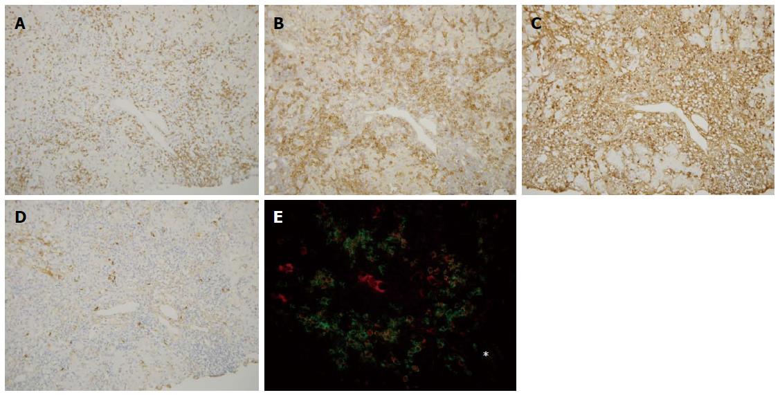
Figure 4 Immunohistochemical findings of autoimmune hepatitis.
CD3+ cells were found within the portal tract and in the periportal area (A), whereas CD38+ cells infiltrated prominently in the periportal area (B). IgG+ cells (C) and IgM+ cells (D) were also found in the periportal area, and IgG+ cell infiltration was predominant in the majority of autoimmune hepatitis cases. The distribution of these cells was similar to primary biliary cirrhosis (Figure 3); E: Double staining for IgG (red) and CD38 (green) showed that IgG-CD38 double positive cells, which may be IgG-producing plasma cells, were frequently seen in the periportal area (star). Original magnifications: × 200. Asterisk: Hepatic lobule.
- Citation: Kobayashi M, Kakuda Y, Harada K, Sato Y, Sasaki M, Ikeda H, Terada M, Mukai M, Kaneko S, Nakanuma Y. Clinicopathological study of primary biliary cirrhosis with interface hepatitis compared to autoimmune hepatitis. World J Gastroenterol 2014; 20(13): 3597-3608
- URL: https://www.wjgnet.com/1007-9327/full/v20/i13/3597.htm
- DOI: https://dx.doi.org/10.3748/wjg.v20.i13.3597


