Copyright
©2013 Baishideng Publishing Group Co.
World J Gastroenterol. Jun 7, 2013; 19(21): 3173-3188
Published online Jun 7, 2013. doi: 10.3748/wjg.v19.i21.3173
Published online Jun 7, 2013. doi: 10.3748/wjg.v19.i21.3173
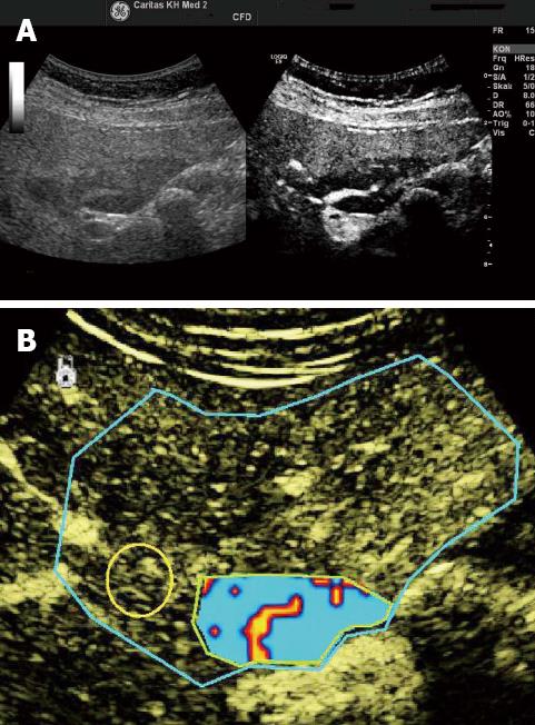
Figure 5 Focal fatty sparing.
A: Focal fatty changes may simulate masses on conventional B-mode ultrasound; B: In the arterial, portal venous and late phases, focal fatty changes show similar enhancement patterns to that of the adjacent liver parenchyma. Contrast-enhanced ultrasound is helpful for the identification of the centrally located arteries. Typically centrally located arteries (and often also portal venous branches and hepatic veins) can be identified. Dynamic vascular pattern improves contrast imaging. Reproduced with permission from Cui et al[120].
- Citation: Dietrich CF, Sharma M, Gibson RN, Schreiber-Dietrich D, Jenssen C. Fortuitously discovered liver lesions. World J Gastroenterol 2013; 19(21): 3173-3188
- URL: https://www.wjgnet.com/1007-9327/full/v19/i21/3173.htm
- DOI: https://dx.doi.org/10.3748/wjg.v19.i21.3173


