Copyright
©2013 Baishideng Publishing Group Co.
World J Gastroenterol. Jan 14, 2013; 19(2): 209-218
Published online Jan 14, 2013. doi: 10.3748/wjg.v19.i2.209
Published online Jan 14, 2013. doi: 10.3748/wjg.v19.i2.209
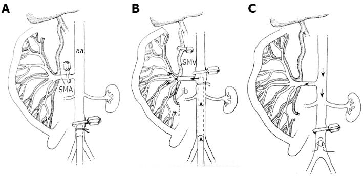
Figure 1 The sequential steps of the intestinal perfusion in vivo.
A: The abdominal aorta (aa) is occluded by a clip, followed by cannulation of the vessel; the superior mesenteric artery (SMA) is clipped; B: The aortic clip is positioned above the SMA; the cannula is positioned at the level of SMA to allow its perfusion with hypertonic saline; the perfusate outflow is secured through the opened ileocecal branch (ib) of the superior mesenteric vein (SMV); the main trunk of SMV is clipped; C: The cannula is removed to allow blood flow through the SMA to resume simultaneously with the following procedures: the aortic clip is positioned as in Figure 1A; the clip occluding the SMV is removed, and bleeding from the ileocecal branch of the SMV is prevented by ligation.
- Citation: Kornyushin O, Galagudza M, Kotslova A, Nutfullina G, Shved N, Nevorotin A, Sedov V, Vlasov T. Intestinal injury can be reduced by intra-arterial postischemic perfusion with hypertonic saline. World J Gastroenterol 2013; 19(2): 209-218
- URL: https://www.wjgnet.com/1007-9327/full/v19/i2/209.htm
- DOI: https://dx.doi.org/10.3748/wjg.v19.i2.209


