Copyright
©2013 Baishideng Publishing Group Co.
World J Gastroenterol. May 7, 2013; 19(17): 2697-2703
Published online May 7, 2013. doi: 10.3748/wjg.v19.i17.2697
Published online May 7, 2013. doi: 10.3748/wjg.v19.i17.2697
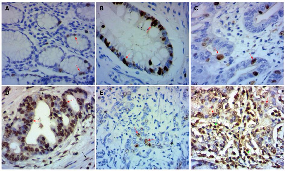
Figure 1 Immunohistochemical staining for distal-less homeobox 2 in gastric adenocarcinoma tissues and adjacent normal gastric tissues.
A: Low expression of distal-less (DLX2) in normal gastric mucosa; B: High expression of DLX2 in intestinal metaplasia cells; C: Low expression of DLX2 in gastric adenocarcinoma tissue with well differentiation; D: High expression of DLX2 in gastric adenocarcinoma tissue with well differentiation; E: Low expression of DLX2 in gastric adenocarcinoma tissue with poor differentiation; F: High expression of DLX2 in gastric adenocarcinoma tissue with poor differentiation. DLX2 staining was detected mainly in nucleus of normal gastric epithelial cells (Figure 1A, red arrows) or tumor cells (Figure 1C-F, red arrows). Besides, increased expression of DLX2 was detected in intestinal metaplasia cells (Figure 1B, red arrows), fibroblasts (Figure 1D, black arrows) and inflammatory cells (Figure 1F, green arrows) around tumor cells. Original magnification, × 200.
- Citation: Tang P, Huang H, Chang J, Zhao GF, Lu ML, Wang Y. Increased expression of DLX2 correlates with advanced stage of gastric adenocarcinoma. World J Gastroenterol 2013; 19(17): 2697-2703
- URL: https://www.wjgnet.com/1007-9327/full/v19/i17/2697.htm
- DOI: https://dx.doi.org/10.3748/wjg.v19.i17.2697


