Copyright
©2012 Baishideng Publishing Group Co.
World J Gastroenterol. Jul 21, 2012; 18(27): 3623-3626
Published online Jul 21, 2012. doi: 10.3748/wjg.v18.i27.3623
Published online Jul 21, 2012. doi: 10.3748/wjg.v18.i27.3623
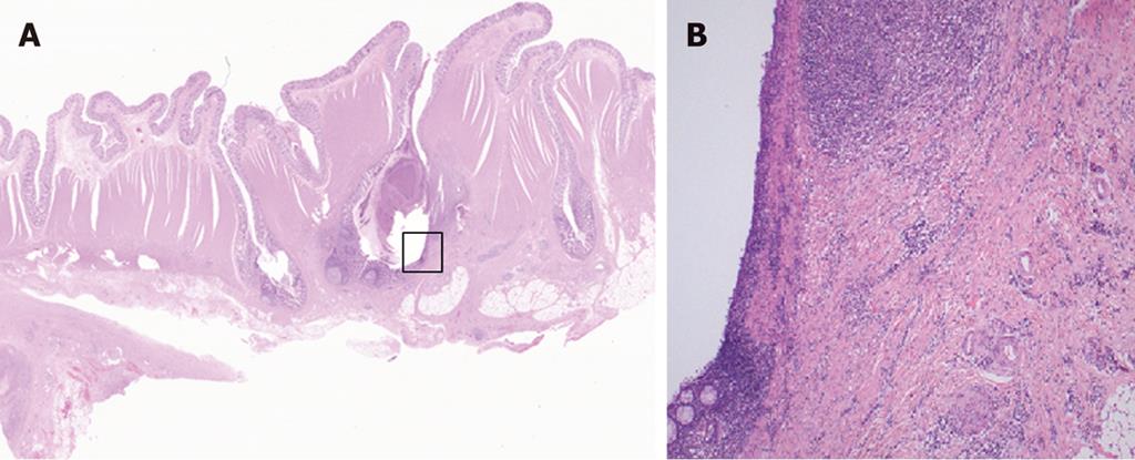
Figure 3 Magnified pathological image of resected specimen.
A: A magnified pathological image of the resected specimen reveals some diverticula in the sigmoid colon (HE stain, 10 ×); B: A magnified pathological image of the marked square in Figure 4A (diverticulosis) shows active inflammation characterized by a marked thickening of the muscularis propria, fibrous thickening of interstitial tissue, and a marked infiltration of inflammatory cells with no evidence of malignancy (100 ×).
- Citation: Nishiyama N, Mori H, Kobara H, Rafiq K, Fujihara S, Kobayashi M, Masaki T. Difficulty in differentiating two cases of sigmoid stenosis by diverticulitis from cancer. World J Gastroenterol 2012; 18(27): 3623-3626
- URL: https://www.wjgnet.com/1007-9327/full/v18/i27/3623.htm
- DOI: https://dx.doi.org/10.3748/wjg.v18.i27.3623


