Copyright
©2011 Baishideng Publishing Group Co.
World J Gastroenterol. May 21, 2011; 17(19): 2357-2364
Published online May 21, 2011. doi: 10.3748/wjg.v17.i19.2357
Published online May 21, 2011. doi: 10.3748/wjg.v17.i19.2357
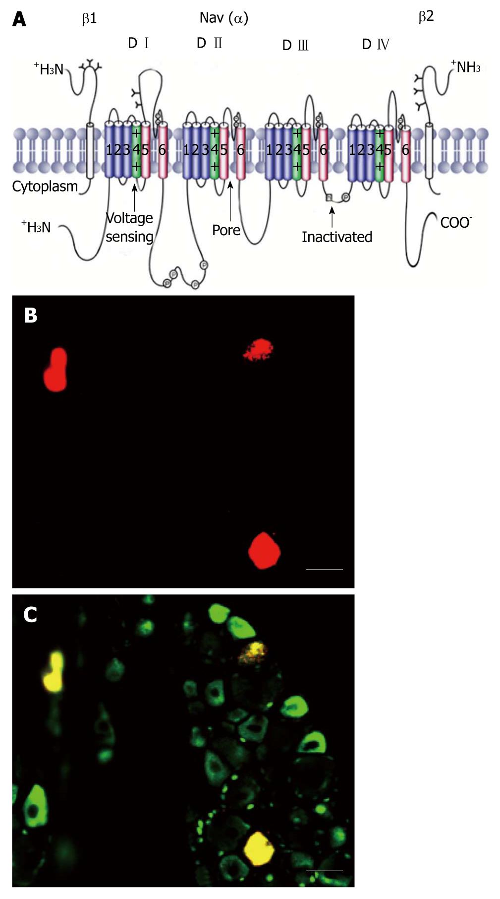
Figure 1 Schematic of a voltage-gated sodium channel polypeptide and Nav1.
8 positive colon specific dorsal root ganglion neurons. A: The transmembrane organization includes four domains (D I-D IV) joined by three intracellular loops (L1-L3) and the intracellular N and C termini. Cylinders represent probable α-helical segments. P in circles, sites of demonstrated protein phosphorylation by protein kinase A (PKA) or PKC; green, S4 voltage sensors; h in which square, inactivation particle in the inactivation gate loop (modified, with permission, from Catterall[14]); B: Colon specific dorsal root ganglion neurons labeled by Dil (red); C: Nav1.8 positive neurons (green). Colon specific Nav1.8 positive neurons are stained yellow. Bar = 50 μm for both B and C.
- Citation: Qi FH, Zhou YL, Xu GY. Targeting voltage-gated sodium channels for treatment for chronic visceral pain. World J Gastroenterol 2011; 17(19): 2357-2364
- URL: https://www.wjgnet.com/1007-9327/full/v17/i19/2357.htm
- DOI: https://dx.doi.org/10.3748/wjg.v17.i19.2357


