Copyright
©2011 Baishideng Publishing Group Co.
World J Gastroenterol. Jan 7, 2011; 17(1): 53-62
Published online Jan 7, 2011. doi: 10.3748/wjg.v17.i1.53
Published online Jan 7, 2011. doi: 10.3748/wjg.v17.i1.53
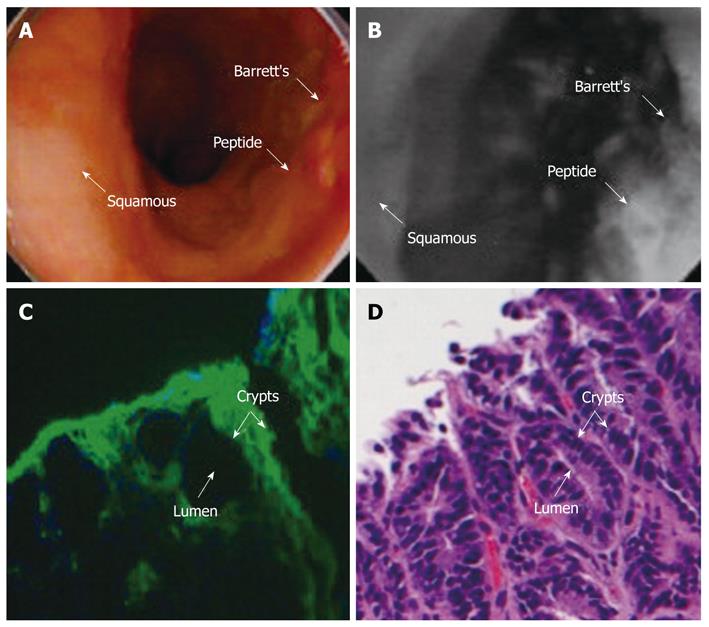
Figure 4 In vivo localization of contrast agent localized to a neoplasia region visualized using wide-field fluorescence endoscopy.
White light endoscopic image (A) shows no evidence of lesion; topical administration of peptide-targeted fluorescent dye reveals neoplastic area (B) [Copyright (2008), with permission from IOS Press][56]; targeted neoplastic crypts seen with fluorescence microscopy (C), and corresponding histology (D) [Copyright (2010), with permission from Elsevier][59].
- Citation: Thekkek N, Anandasabapathy S, Richards-Kortum R. Optical molecular imaging for detection of Barrett’s-associated neoplasia. World J Gastroenterol 2011; 17(1): 53-62
- URL: https://www.wjgnet.com/1007-9327/full/v17/i1/53.htm
- DOI: https://dx.doi.org/10.3748/wjg.v17.i1.53


